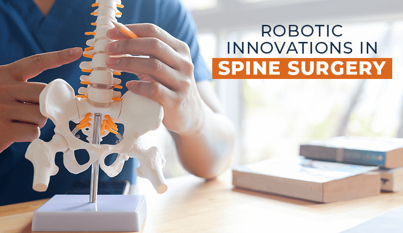
Robotic Spinal Surgery and the Mazor X System

Over the past few decades, the field of spinal surgery has witnessed significant transformative developments. From the early days of free-hand techniques to the development of modern, image-guided approaches using fluoroscopy and intraoperative CT scans, each phase brought greater precision and safety to the operating room.
Technological advances in spinal surgery include the use of robotic platforms that aim to enhance surgical accuracy and consistency. The Mazor X system, for example, integrates imaging and software guidance to assist surgeons with preoperative planning and intraoperative control.
As the demand for minimally invasive spine surgery grows - driven by the desire for quicker recovery times, reduced postoperative pain, and lower complication rates - robotic systems are becoming increasingly prominent. They promise to transform patient experiences, surgeon workflows, and healthcare outcomes, making surgeries safer, smarter, and more efficient.
This article examines the evolution of spine surgical techniques, the emergence of robotics, conditions treated, challenges encountered, and future directions.
What is Robotic Spinal Surgery?
Robotic spinal surgery, also referred to as robot-assisted spinal surgery, is a surgical technique that integrates robotic technology with real-time imaging and computer-assisted navigation to achieve enhanced precision and safety in spinal procedures. These systems act as extensions of the surgeon’s expertise.
In robotic-assisted spinal surgery, surgeons can pre-plan each step of the operation using high-resolution 3D imaging. This data is fed into the robotic system, which then guides the surgical instruments with millimeter-level accuracy to the predetermined anatomical targets. The result is greater precision in procedures like pedicle screw placement, spinal fusions, and correction of spinal deformities.
There are three primary types of robotic surgical systems used across various medical disciplines, including spine surgery:
- Shared-Control Systems: In this setup, the surgeon and robot work collaboratively. The surgeon retains full control, but the robotic system offers resistance, stabilization, or guidance to ensure precise movement within predefined boundaries. These systems help reduce human error and enhance instrument stability.
- Supervisory-Controlled Robots: These robots execute specific tasks that have been pre-programmed and pre-planned by the surgeon. Once the plan is finalized, the robot operates autonomously under supervision, typically performing tasks such as bone milling, screw insertion, or tissue retraction with consistent accuracy.
- Telesurgical Robots: Often associated with systems like the da Vinci Surgical System, telesurgical robots allow surgeons to operate remotely using a console. While this approach is less common in spine surgery, advances in robotic telepresence may eventually enable spinal procedures to be performed remotely, particularly in underserved or remote regions.
At present, shared-control systems are the most commonly implemented robotic surgical systems in the field of orthopedics and spine care.
The Evolution From Free-Hand to Robotics
Free‑Hand Technique: Before the adoption of image guidance, spinal surgeries relied primarily on the free-hand technique. Surgeons depended on anatomical landmarks and tactile feedback to place pedicle screws and other instrumentation. This approach required extensive exposure of bony anatomy, often necessitating large incisions, and was prone to higher rates of misplacement in complex cases.
Fluoroscopy‑Guided 2D Navigation: To improve safety and accuracy, image-guided techniques were introduced in the 1990s. Early methods employed C‑arm fluoroscopy for 2D guidance during screw insertion. While fluoroscopy enhanced visualization compared to blind techniques, it still relied on the surgeon’s hand-eye coordination and involved significant radiation exposure to the surgical team.
Intraoperative 3D CT Navigation: The next major leap was the introduction of intraoperative 3D imaging using mobile CT devices, such as the O-arm, as well as surgical navigation systems. These systems registered patient anatomy using preoperative or intraoperative CT data, enabling real-time instrument tracking in true 3D space. Incorporating navigation improved screw placement accuracy and reduced radiation exposure, while preserving the surgeon’s tactile control over instruments.
Robotic‑Assisted Systems: Integrating planning, navigation, and robot guidance, exemplified by systems like Mazor X, marking the latest phase in image-gated spine surgery.
About the Mazor X Robotic Guidance System
System Type
The Mazor X is a shared-control robotic guidance system, a collaborative platform that augments the surgeon's manual skills rather than replacing them. It integrates preoperative planning, robotic arm guidance, and real-time navigation, enabling the surgeon to retain control while the robot ensures precise trajectory adherence.
Key Features of the Mazor X System
-
3D Preoperative Planning
Utilizing CT-based images, Mazor X automatically recognizes spinal anatomy to build 3D models. Surgeons can simulate screw trajectories, optimize rod and cage placement, plan skin incisions, osteotomies, and tailor implant selections, all in a virtual environment before entering the OR.
-
Robotic Arm with Guidance Technology
On surgery day, the robotic arm locks to the bed-mounted platform, establishing a rigid connection with the patient’s spine. It positions tools along the pre-planned trajectories with sub-millimeter precision, reducing human deviation and enabling efficient screw placement, even in complex anatomical scenarios or revision cases.
-
Real-time Intra‑operative Navigation Integration
Mazor X stealth edition integrates with Medtronic’s StealthStation navigation. Live tracking enables visualization of instruments in relation to the patient’s anatomy and planned paths, completing the loop between planning and execution and providing depth monitoring alongside robotic guidance.
-
Guidance Modules and Attachments
Dedicated attachments, such as the Midas Rex high-speed drill, tap guides, and screwdriver systems, ensure that drill and screw paths adhere precisely to the robotic arm’s trajectory. These tools often integrate optical trackers to maintain navigational accuracy during instrumentation.
-
Predictive Analytics & 3D Analytics
The integrated planning software provides detailed 3D analysis to evaluate design and alignment, pedicle screw placement, osteotomy requirements, and load distribution. This allows surgeons to anticipate anatomical challenges and tailor fusion strategies to the patient’s specific condition and needs.
Workflow: From Pre‑planning to Recovery
Pre‑planning
- The patient undergoes a high-resolution CT scan.
- The surgical team constructs a 3D model and simulates ideal trajectories, selecting implants, rod offsets, and incision sites.
- The construct design and global spinal alignment are reviewed and optimized using analytics.
Setup & Registration
- On OR day, the patient and the robotic arm are rigidly secured via a bed-mounted frame.
- CT images are registered to intraoperative fluoroscopy (or O-arm) scans to ensure accurate alignment with the patient’s actual anatomy and the preoperative plan.
Procedure Execution
- The robotic arm positions guiding cannulas. Instruments are introduced through these guides, ensuring accuracy.
- Real-time navigation and periodic imaging confirm instrument placement depth and orientation.
- Procedures such as pedicle screw placement, decompression, transforaminal lumbar interbody fusion (TLIF), or retractor placement proceed with robotic support.
Recovery
- Robotic assistance minimizes soft tissue damage, optimizes accuracy, and reduces radiation exposure.
- Resulting benefits include less intraoperative blood loss, shorter anesthesia times, faster recovery, and smaller incisions, leading to shorter hospital stays.
Common Procedures Performed Using Mazor X
- Thoracolumbar spinal fusion: Robotic navigation supports accurate screw placement and optimal spinal alignment for fusion procedures.
- Scoliosis and deformity correction, including kyphosis and spondylolisthesis cases: Used to plan spinal realignment and guide precise correction of complex deformities.
- Vertebral body tethering: Helps ensure safe, accurate tether placement to modulate spinal growth while preserving motion.
- Pedicle screw fixation: Improves accuracy and consistency in planning and inserting pedicle screws.
- Degenerative disc disease management, including herniated discs and TLIF: Assists in disc access, implant positioning, and spinal alignment during minimally invasive procedures.
- Trauma and tumor pathology, including fracture fixation and tumor resection: Aids in stabilizing fractures and guiding precise resections near delicate spinal structures.
How Mazor X Enhances Spinal Surgery
Robotic assistance via the Mazor X system significantly advances the precision, safety, and outcomes of spinal procedures across multiple dimensions.
Improved Screw Placement Accuracy
According to published literature, robotic systems, such as the Mazor X, have consistently demonstrated accuracy rates of 98% (and higher) in pedicle screw placement. This level of precision far exceeds traditional free-hand techniques, which carry a higher risk of misplacement. The robotic arm and navigation technology work together to ensure the implants are placed precisely as planned.
Minimally Invasive Capabilities
Mazor X enables minimally invasive spine surgery by guiding instruments through smaller incisions and preserving muscle tissue. Consequently, patients experience reduced postoperative pain, less blood loss, and shorter hospital stays. Minimally invasiveness also reduces the risk of infection and enables faster recovery times.
Complex Deformity Correction
The correction of complex spinal deformities such as scoliosis, kyphosis, and spondylolisthesis can particularly benefit from the system. Mazor X allows surgeons to map the spinal column in detail and plan optimal correction strategies tailored to each patient’s anatomy.
Personalized Surgical Planning
Through preoperative CT-based imaging and planning software, surgeons can design a surgical plan customized to each patient’s spinal anatomy. They can choose optimal trajectories for screw placement and simulate the procedure ahead of time, which helps avoid intraoperative surprises.
Reduction in Radiation Exposure
By decreasing the number of intraoperative fluoroscopic images needed, Mazor X reduces radiation exposure for both patients and surgical staff. This is a significant benefit over conventional techniques, where continuous imaging is often required for alignment verification.
Lower Risk of Complications or Revision Surgery
The combination of accurate screw placement and personalized planning reduces the likelihood of surgical complications, hardware failure, or the need for reoperations. This helps improve long-term outcomes and patient satisfaction.
Enhanced Precision and Control
Mazor X reduces hand tremor, enabling a stable and controlled environment for guiding instruments. The robotic arm operates with sub-millimeter precision, allowing surgeons to work in delicate areas of the spine with confidence.
Reduced Physical Strain for Surgeons
Robotic systems ease the physical burden on surgeons by supporting instrument positioning and maintaining a steady hand during long procedures. This can reduce fatigue and improve performance during complex operations.
Near-Zero Implant Misplacement
When used appropriately, Mazor X offers near-zero rates of implant misplacement. This outcome is critical in avoiding neurological injury and ensuring that spinal hardware performs as intended.
Effectiveness in Revision Surgeries
Mazor X is highly effective in revision spine surgeries. When previous procedures have failed or hardware needs to be corrected or replaced, the system provides precise visualization and navigation to manage scar tissue and anatomical changes.
Challenges and Considerations
Despite its benefits, Mazor X is not without limitations.
High Initial Cost and Training Curve
Mazor X requires a significant financial investment and specialized training. It is typically available only in large hospitals or advanced medical centers, which, from the patients’ point of view, means that it is not widely accessible.
Not Suitable for All Patients
There are contraindications where Mazor X may not be appropriate, such as in cases of severe cervical instability or highly atypical anatomy (e.g., severe obesity, osteoporosis). Patient selection remains an important factor in its success.
Dependence on Imaging Quality and Preoperative Planning
The system relies heavily on the accuracy of preoperative imaging. Any errors in CT scans or planning can affect the outcome. Meticulous preparation is essential to ensure that the robotic guidance matches the patient’s anatomy during surgery.
Potential for Technical Issues
As with any advanced technology, there is a risk of technical malfunctions or software errors. Backup protocols must be in place, and the surgical team must be prepared to revert to manual techniques if necessary.
The Future of Robotic Spine Surgery
The future of robotic spine surgery is poised for further innovation.
Integration with Artificial Intelligence
Emerging systems may integrate AI to analyze anatomical data and suggest optimal surgical plans. AI could also help adapt the robotic system in real time during the procedure, improving decision-making and outcomes.
Real-Time Intraoperative Adaptability
Next-generation robots may respond to anatomical changes or surgical variables during the operation. This dynamic feedback loop could further enhance accuracy and safety.
Expanding Indications and Applications
While Mazor X currently focuses on thoracolumbar procedures, future advancements may expand its use into cervical spine surgeries, motion-preserving techniques, and more outpatient or same-day procedures.
Innovative Systems
Several robotic platforms are also under development or in clinical use. These include ExcelsiusGPS by Globus Medical, ROSA Spine by Zimmer Biomet, Fusion Robotics, CURVE by DePuy Synthes, and others. Each system offers distinct features and approaches, pushing the field forward through competition and innovation.

Dr. Fernando Álvarez Sala-Walther is a leading orthopedic spine surgeon and the Head of the Spinal Pathology Unit at Hospital Ruber Internacional from Quirónsalud Hospital Group. He is internationally recognized for his expertise in robotic and image-guided spinal surgery. Dr. Álvarez Sala-Walther combines cutting-edge technology with personalized care to treat complex spinal conditions, including deformities, degenerative diseases, trauma, and tumors. He is actively involved in clinical research and has contributed to the advancement of precision spine surgery in Spain.

Hospital Ruber Internacional from Quirónsalud Hospital Group is a leading private hospital in Madrid, Spain, known for its top specialists, advanced technology, focus on innovation, and commitment to continual advancement in healthcare. The hospital offers cutting-edge treatments in various specialties, such as cancer, cardiovascular, and neurological conditions.
The Spinal Pathology Unit at Hospital Ruber Internacional contributes to ongoing advancements in spinal care through its clinical expertise in managing degenerative, traumatic, and complex spinal conditions. The hospital serves as a reference center for robotic-assisted spine surgery in Spain, supporting evidence-based approaches and integrating emerging technologies in line with current surgical standards.
Reference:
Featured Blogs


