Cardiovascular System Overview

Medically Reviewed by Dr. Sony Sherpa, (MBBS) - September 13, 2024
“The art of medicine has its roots in the heart.
If your heart is false, then also the doctor in you is false.
If it is fair, then also the doctor is fair.”
- Paracelsus
As a major component of our physiology as complex organisms, understanding the cardiovascular system is an important step to take if one wants to be an advocate for one’s own health and well-being. Below, the components that complete the cardiovascular system are discussed, as well as their main functions and how they work together to promote balance at all times.
Main Functions of the Cardiovascular System
When thinking about what it means to be alive and well, the cardiovascular system is one of the main areas of the body that first comes to mind. Often, vitality, love, passion, energy, hard work, and, to some of us, the purity of spirit have been interwoven with the concept of the blood that flows through our veins. The heartbeat unifies us, being a common denominator among all human beings, and takes us back to the time we spent in utero, synonymous with the origins of life and creation.
The functions of the cardiovascular system emulate these abstract connotations. Blood is responsible for ensuring that every tissue remains oxygenated and capable of producing energy. It also takes waste products away from every tissue, maintaining a literal biological balance between creation and destruction.
Aside from being the primary transport system of oxygen and carbon dioxide, the cardiovascular system also ensures that vital nutrients, hazardous toxins, white blood cells, and important cell signals (e.g., inflammatory compounds and hormones) reach their desired destinations. Blood vessels also serve to regulate the pH of the body, body temperature, blood pressure, and overall osmotic pressure of bodily fluids.
Without these functions of the vascular system, every other cell in the body would cease to function properly due to poor maintenance and rapid deterioration. In light of this, the vascular system is as important as our nervous system in terms of the role it plays in generating our lived experience and sustaining our biological existence.
The Working Parts of the Cardiovascular System
The cardiovascular system mainly comprises the blood, blood vessels, and the heart, with some involvement from the nervous system, immune system, and digestive system.
Blood
Blood is the only fluid considered to be a tissue in the body, making it the most flexible and adaptive bodily “organ.” It consists of plasma, the liquid base that houses red blood cells and proteins, which together make up blood as a whole. Plasma and plasma proteins proportionally comprise 55% of the blood, while red blood cells make up the other 45%.
The proteins in the blood are largely produced by the liver, bone marrow, and spleen, with contributions also from dead blood cells and the cells (endothelial cells) that comprise our blood vessels (the endothelium). Blood plasma itself is created from water and mineral salts, extracted and replenished during digestion. In a person weighing an average of 70kg, extracellular fluids consist of ±14L (±20% of total body weight). The fluids inside our cells, by contrast, account for roughly double that amount (±40% of total body weight).
The most important constituents of blood include[1]:
- Albumins and Globulins. These are the two main types of plasma proteins found in circulation. All types are involved in transporting hormones, ionic (charged) particles, vitamins, toxins, and many other kinds of proteins around the body. The majority of chemical compounds that remain unbound in blood plasma are potentially harmful to the body. Immunoglobulins are synthesized by white blood cells (B lymphocytes) and are designed to chaperone foreign proteins that pose threats to security (an antibody binding to an antigen), as well as participate in allergic reactions to allergens. Certain globulins are produced to bind to hormones, such as sex hormone-binding globulin[2] and corticosteroid-binding globulin[3], and chaperoning sex hormones like estrogen and glucocorticoids (stress chemicals), respectively. Albumins bind to a wide variety of substances, including free hormones and calcium molecules. It also enters the spaces between cells when an area becomes microscopically damaged or when membranes become permeable, in order to prevent spillage from both macro and micro bodily compartments.
- Unbound Chemicals, Toxins, Cell Debris and Pathogens. These are substances in the blood that are potentially harmful and often engage the immune system wing present in the bloodstream. The liver does its best to prevent toxins that we eat from entering systemic circulation; however, when there is a breach in the lining of the gut wall, many toxins can enter hepatic (liver) circulation and overburden the organ, resulting in transient systemic toxicity.
- Electrolytes. Electrolytes, namely sodium, calcium, potassium, chloride, magnesium, and other relevant substances like bicarbonate, all help to maintain optimal blood pressure and pH. Many of these nutrients are also required by all cells in order to generate energy and are appropriately transported to all cells via the bloodstream.
- Coagulants. Coagulants consist of other types of proteins that collectively clot blood in order to facilitate wound healing. Blood is one of the most vulnerable tissues when exposed to the outer environment. Thus, blood clotting factors help prevent blood loss and eventually serve as a temporary membrane for this fluid organ until the wound is repaired.
- Red Blood Cells. Red blood cells play a vital role in transporting oxygen around the body and removing carbon dioxide from the body. The protein in blood cells that makes them red is known as hemoglobin, the iron-rich heme portion of the molecule responsible for this coloration and the binding of oxygen. Oxygen requires this kind of specialized transport as it is a highly reactive substance. The mitochondria in every cell receive oxygen, and through a very careful set of chemically controlled steps, they process it into tangible biological energy.
- White Blood Cells. White blood cells also patrol the bloodstream. The main ones active, while there is no apparent infection or other threat, include phagocytes such as macrophages and B lymphocytes such as basophils and eosinophils. The former is involved in identifying threats and consuming potentially hazardous material in order to break it down into harmless compounds to be eliminated. The latter are involved in identifying allergens or foreign proteins that are out of place, which may trigger an allergic response.
The Heart
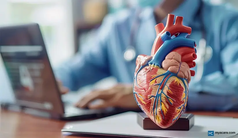
The heart is the central processing unit for the cardiovascular system in much the same way that the brain is for the nervous system. It sustains the movement of blood through tirelessly relaxing and contracting, which is essentially the essence of our heartbeat.
The Heart Beat
The cells that make up the heart are known as myocytes, and each one is packed with thousands of mitochondria to sustain the energy required to keep the heart beating all the time. This mechanism is also governed by the electrolyte balance in the bloodstream and in myocytes, with sodium and potassium forming the main axis upon which fluids circulate inside the heart in order to achieve constant contractions. This is sometimes referred to as the sodium-potassium pump or the Na/K-ATPase pump.
The Chambers of the Heart
The heart is divided into four chambers that contain valves in order to prevent the backflow of blood, keeping it moving in one continuous direction. The side of the heart to our left, the side where the heart is positioned in the body, consists of the left atrium and left ventricle, while the right-hand side contains the right atrium and right ventricle. Both left and right atria rest above the left and right ventricles, respectively.
The left side of the heart processes oxygenated blood, while the right-hand side processes deoxygenated blood. Blood starts off being oxygenated when expelled from the heart and ends up being deoxygenated by the time it returns. In the lungs and in the periphery of the body, the veins and arteries meet in order to complete circulation and allow for deoxygenated blood to become oxygenated again.[4]
Blood Vessels
The blood vessels are basically an extension of the heart, emanating out from the heart like branches from a tree. The thicker blood vessels comprise multiple layers, some of which are composed of smooth muscle tissue that is able to contract like the muscle tissue of the heart, aiding blood flow.
The main types of blood vessels are as follows:
Arteries
Arteries carry oxygenated blood away from the heart. The aorta is the main artery and is also the thickest blood vessel. Freshly oxygenated blood from the lungs enters the heart via the left atrium before being expelled out to the rest of the body via the left ventricle and into the aortic artery. The aorta branches off into smaller arteries that feed to the periphery and into all other organs and tissues throughout the body. In organs and peripheral tissues, this blood loses its oxygen content before making its way back toward the heart through the veins.
Veins
The veins carry deoxygenated blood to the heart. In general, blood pressure is lower in veins than it is in arteries. As with the arteries, the veins are thickest around the heart and get thinner as they branch out. The thickest vein is known as the vena cava. It is directly connected to the heart, and eventually, all deoxygenated blood travels through it into the right atrium. From there, it travels through to the right ventricle and out toward the lungs, where the blood cells exchange carbon dioxide for more oxygen. The cycle completes when this newly oxygenated blood travels back to the heart and is used once more to oxygenate all bodily tissues.
Capillaries
Capillaries are the smallest type of blood vessels and are also where the veins and arteries of the body meet in order to complete the circuit. These minute blood vessels are so small that only one red blood cell may pass through at a time. In most cases, blood cells have the natural ability to be flexible, and in the smallest of these vascular spaces, they need to bend in order to squeeze through. This narrow passage helps to maintain optimal blood pressure each time the blood is pumped from the larger veins surrounding the heart. If the blood vessels were all one size, blood would lose momentum and be unable to reach all parts of the body.
Nervous System
The nervous system helps to maintain optimal functionality in the heart at all times. There are a number of different neuro-receptors found in the heart and its surrounding vascular tissues. The two main types consist of baroreceptors and chemoreceptors.[5]
Baroreceptors
Baroreceptors respond to changes in blood pressure, sending sensory information back to the brain when pressure increases or decreases in the aorta and heart valves. For normal heart function, baroreceptors need to be stretched by the pressure in the bloodstream. Too little pressure coupled with less sensory feedback causes the brain to increase activity in the sympathetic nervous system and initiate a stress response. This, in turn, causes blood vessels to constrict via smooth muscle contraction in order to boost blood pressure back into a normal range again. The opposite process is true of high blood pressure.
Peripheral Chemoreceptors
Peripheral Chemoreceptors are the first kind of chemoreceptor. Like baroreceptors, these are also located in the aorta and in nearby cardiac tissues. Peripheral chemoreceptors don’t respond to physical stimulation but instead respond to chemical changes, such as fluctuations in oxygen, carbon dioxide, and pH levels. When oxygen is too low, carbon dioxide too high, and pH too acidic, chemoreceptors become active and send signals back to the brain. Depending on other factors, the brain can respond in a variety of ways that ensure more oxygen enters circulation.
Central Chemoreceptors
Central Chemoreceptors are found in the brain stem (specifically the medulla oblongata), and just like those found in the heart and blood vessels, these receptors also respond to changes in pH, oxygen, and carbon dioxide. Their specific focus is on the blood that oxygenates cerebrospinal fluid, which helps to monitor the state of oxygenation in the brain.
Vagus Nerve
The vagus nerve also connects to the heart and plays a role in modulating cardiac output during digestion and relaxation (parasympathetic nervous system activity), as well as during acute stress (sympathetic nervous system activity). Consuming food that is too acidic without a balancing component, such as excessively fatty foods with low nutritional content, tends to cause gastric reflux as well as possible heart palpitations or changes in the rhythm of the heartbeat. Indeed, gastric and other digestive symptoms can initiate a stress response that indirectly raises the heart rate, or they can directly stimulate the heart via the vagus nerve. In this way, the heart is connected to the digestive system.
Cardiovascular Division of Blood Flow
On top of the way that the heart and blood vessels separate out oxygenated and deoxygenated blood, additional compartments of organization reside within our vasculature. There are two main circuits for blood to flow throughout the body that are governed exclusively by the cardiovascular system: pulmonary circulation and systemic circulation.
Some organs have their own unique circuits within the systemic one, but those smaller circuits are still powered by the same mechanisms that manage systemic circulation in its entirety.
Pulmonary Circulation
Pulmonary circulation refers to the veins and arteries that connect the heart to the lungs. The prime function of this circuit is to take deoxygenated blood and re-oxygenate it. This is achieved via gaseous exchange in the incredibly fine capillary network residing in the alveoli of the lungs.
When deoxygenated blood from the body enters the heart, it fills up the right atrium at the same time that oxygenated blood from the lungs fills up the left atrium. The atria form the top half of the heart.
Before the heart contracts, it relaxes, allowing for this blood to move into the left and right ventricles respectively – the bottom half of the heart. When the heart contracts, this blood is ejected from the heart, with newly oxygenated blood from the left ventricle passing into systemic circulation and deoxygenated blood coming from systemic circulation moving over into pulmonary circulation.
Systemic Circulation
Systemic circulation refers to the blood vessels that form a circuit between the heart and the remainder of the body. As described above, the heart ejects oxygenated blood, freshly renewed from pulmonary circulation, into the rest of the body. At the same time, deoxygenated blood is pushed back toward the heart to make its way into pulmonary circulation for more oxygen.
Another critical difference between systemic and pulmonary circulation is that systemic circulation is closed off from the environment, while pulmonary circulation is made more vulnerable via the lungs and airways. Systemic circulation is more involved in transporting nutrients throughout the body as a whole, while pulmonary circulation mostly maintains the oxygenation of blood as well as nutrient transfer in the respiratory system.
Organ-Specific Blood Barriers
Naturally, every organ and nearly all tissues are connected by the cardiovascular system. Some organs have specialized filtration systems that form structural parts of our overall circulation. The liver, with its hepatic portal vein, and the brain, with its blood-brain barrier, are the two most important examples:
- Hepatic Portal Vein. The liver filters out toxins from nutrients that are directly absorbed from the stomach, either allowing or preventing them from entering circulation. Beyond the realm of the stomach, this barrier to circulation in the liver monitors all nutrients that have been absorbed during digestion, allowing only safe particles to pass into systemic circulation. Anything that is rejected tends to traverse via the gallbladder and be emptied back into the digestive tract for fecal excretion or is chemically bound and allowed to travel through the blood toward the kidneys or skin for other types of excretion.
- Blood-Brain Barrier. The brain is a very sensitive organ that requires optimal blood flow and as little toxin interference as possible to function at its optimum. The blood-brain barrier ensures that the majority of substances in the bloodstream cannot pass into the cranium, keeping the environment in there as pristine as possible. This is vital to our functioning as the brain is only capable of cleansing itself when we sleep, generating its own load of toxins throughout the day.
Autoregulation of Blood Flow
Autoregulation is an official term given to the mechanisms that ensure all organs of the body receive adequate blood flow[6]. When blood flow is restricted to an organ, blood vessels dilate to accommodate more blood flow by reducing resistance to the area.
Scientists are still debating over theories as to how the blood vessels achieve autoregulation, effortlessly maintaining blood flow to all of its tissues. The likeliest answer is that it is a combination of all observable theories, as blood flow is far too important to our basic survival as organisms.
The following theories and observations all form part of the currently accepted understanding of autoregulation.
Myogenic Regulation
The myogenic theory states that blood flow is regulated by the intrinsic properties of smooth muscle in blood vessels. When there is an increase in the exchange of gases (oxygen and carbon dioxide) in the blood, it causes blood vessels to dilate and stretch, which in turn stimulates smooth muscle contractions via the nervous system to keep the blood moving steadily forward. In the opposite case, too little stretching of the vessels causes their smooth muscle tissue to relax, resulting in blood vessel dilation, lower pressure, lower mechanical resistance, and the easy movement of blood.
Metabolic Regulation
The metabolic theory of autoregulation arose from the observation that wherever there is metabolic activity at the cellular level, blood flow is not far behind to aid the process. The metabolic activity referred to here is not related to digestion but to energy production and cellular energy metabolism.
It’s only natural for blood to follow metabolic activity in the body, as oxygen and other nutrients present in the blood are the prime requirements to sustain energy generation.
When energy output is increased in a tissue, the cells in that tissue release gases like nitric oxide, which serve to dilate blood vessels and enhance blood flow. Other by-products released in the process that increase dilation include adenosine, potassium, hydrogen ions, lactic acid, and carbon dioxide. This is why it is a common case that after working out or breaking a sweat, our blood vessels expand and rise to the surface of the skin. It also helps with letting off the extra heat generated in the process of increased energy production.
Organ-Specific Autoregulation
Each organ and tissue has a unique way of maintaining autoregulation. The following highlights what is known about autoregulation in major organs and tissues throughout the body:
- Skin. Blood flow to the skin is regulated via the nervous system and often in response to changes in temperature. Warmer ambient temperatures increase blood vessel dilation as well as keep the blood in a more fluid state. Colder environments cause blood vessels to constrict to facilitate optimal blood flow.
- Muscle. During exercise, autoregulation of muscle appears to be governed by metabolic regulation. In a state of rest, blood flow to muscles is largely controlled by myogenic regulation.
- Brain. The brain’s blood barrier prevents many metabolites that would usually cause metabolic vasodilation, and the blood vessels present in the brain lack the smooth muscles that the ones nearer the heart possess. Instead, blood flow to the brain is largely regulated through carbon dioxide alone, with an increase causing vasodilation and a decrease causing vasoconstriction.
Other waste products are collected at night by the cerebrospinal fluid, which experiences exchange with the bloodstream in the spinal cord. Oxygen, interestingly, can cause vasodilation when it is too low in the brain but does not cause constriction when levels are higher than usual. It seems that the brain uses every bit of oxygen that it can to power itself! This observation is also supportive of the fact that sometimes, all one needs is some fresh air to clear the head.
- Heart. The heart is governed by both types of regulation, with adenosine, nitric oxide, and higher levels of carbon dioxide than oxygen causing vasodilation.
- Lungs. When there is too little oxygen in the lungs, it causes myogenic constriction, which facilitates blood flow to areas where there is an available supply of oxygen.
- Kidneys. The kidneys are regulated largely through nervous myogenic regulation. The filtration tubules that work to collect useful substances from pre-processed urea contribute to autoregulation by stretching, signaling for vasodilation to improve blood flow to the area. Extremely low blood pressure can cause the vessels in the kidney to overly constrict, resulting in renal failure.
To search for the best cardiology healthcare providers in Croatia, Germany, India, Malaysia, Poland, Singapore, Spain, Thailand, Turkey, the UAE, the UK and The USA, please use the Mya Care Search engine.
To search for the best healthcare providers worldwide, please use the Mya Care search engine.
The Mya Care Editorial Team comprises medical doctors and qualified professionals with a background in healthcare, dedicated to delivering trustworthy, evidence-based health content.
Our team draws on authoritative sources, including systematic reviews published in top-tier medical journals, the latest academic and professional books by renowned experts, and official guidelines from authoritative global health organizations. This rigorous process ensures every article reflects current medical standards and is regularly updated to include the latest healthcare insights.

Dr. Sony Sherpa completed her MBBS at Guangzhou Medical University, China. She is a resident doctor, researcher, and medical writer who believes in the importance of accessible, quality healthcare for everyone. Her work in the healthcare field is focused on improving the well-being of individuals and communities, ensuring they receive the necessary care and support for a healthy and fulfilling life.
Sources:
Featured Blogs


