
Understanding Basal Cell Carcinoma: Symptoms, Types & Treatment

Medically Reviewed by Dr. Dora Matis - June 16, 2026
Basal cell carcinoma is a slow-growing skin cancer that usually forms on sun-exposed areas. Basal cell carcinoma has several subtypes, including nodular and superficial forms, and is strongly associated with ultraviolet radiation exposure. Standard diagnostic verification requires dermoscopy and a skin biopsy to differentiate it from look-alikes such as melanoma. Surgical excision, Mohs micrographic surgery (for selected high-risk cases), cryotherapy, and topical medications are some of the treatment options. While early-stage prognosis yields excellent survival rates, neglected cases risk deep, destructive tissue invasion.
Introduction
Globally, skin cancer is one of the most commonly diagnosed malignancies, and its incidence continues to rise in many populations. The main causes of this trend are:
- Increasing life expectancy
- Cumulative sun exposure
- Lifestyle factors
Among skin cancers, non-melanoma basal cell carcinoma accounts for seventy to eighty percent of all new cases, making it the most frequently diagnosed type.
The encouraging news is that the disease is often discovered as early-stage basal cell carcinoma. In such cases, the treatment is usually simple, and outcomes are excellent. However, awareness remains crucial. Neglected lesions may progress and cause significant local damage. Therefore, early detection is important for maintaining both health and physical appearance.
What Is Basal Cell Carcinoma?
Basal cell carcinoma is a type of malignant skin tumor. It develops from basal cells located in the deepest layer of the epidermis. These cells are responsible for continuous skin renewal. Here are some important basal cell carcinoma characteristics:
- Slow but persistent growth
- Local tissue invasion
- Very low metastatic potential
- Strong association with chronic sun exposure
How fast does basal cell carcinoma grow?
Usually, the tumor growth occurs over months to years. That is the key reason why these newly formed lesions are sometimes ignored for too long. This also emphasizes why understanding basal cell carcinoma and how long it takes to grow is crucial for a timely diagnosis.
What Are the Types of Basal Cell Carcinoma?
Basal cell carcinoma has several different subtypes. Here is a quick overview:
-
Nodular Basal Cell Carcinoma
This is the most common subtype. It typically appears as a pearly, dome-shaped lesion with visible blood vessels, frequently developing as basal cell carcinoma on the face, particularly the cheeks, nose, and forehead.
-
Superficial Basal Cell Carcinoma
Usually presents as a thin, red, scaly patch on the trunk or shoulders and may resemble inflammatory skin conditions.
-
Infiltrative and Morpheaform Type
These aggressive subtypes tend to grow with poorly defined borders and a scar-like appearance. They have a higher risk of incomplete excision and recurrence.
-
Pigmented Variant
Contains brown, blue, or black pigmentation and may mimic melanoma.
What Are the Causes and Risk Factors of Basal Cell Carcinoma?
Primarily, ultraviolet radiation from sunlight or tanning beds causes basal cell carcinoma. Additional risk factors include:
- Fair skin, light hair, and light eyes
- Increasing age and male sex
- Personal or family history of skin cancer
- Immunosuppression
- Environmental exposures such as arsenic or radiation
- Genetic conditions like nevoid basal cell carcinoma syndrome
If left untreated, basal cell carcinoma can continue to grow and invade deeper tissues, potentially causing significant local damage and functional impairment.
What Are the Symptoms of Basal Cell Carcinoma?
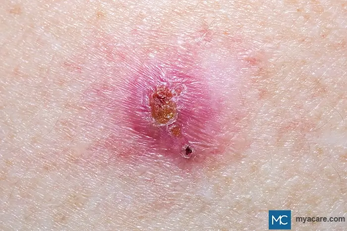
The first obvious sign of basal cell carcinoma is the formation of a skin lesion that can manifest as:
- A shiny or pearly bump
- A non-healing sore
- A flat, red scaly patch
- A firm, scar-like area
These usually appear in sun-exposed areas.
In early-stage 1 basal cell carcinoma, lesions are usually small, asymptomatic, and slow-growing. Understandably, this makes them easy to overlook.
Although distant spreading of the tumor is rare, advanced basal cell carcinoma can invade cartilage, muscle, and bone. Stage-4 basal cell carcinoma is rare. However, it may occur in long-standing or aggressive cases.
Commonly affected locations include the face, scalp, ears, and neck. Additionally, the eyelid may be affected, with basal cell carcinoma of the eyelid representing the majority of eyelid malignancies.
How Is Basal Cell Carcinoma Diagnosed?
Diagnosis begins with clinical examination. This is followed by dermoscopy to identify characteristic vascular and pigment patterns. The final confirmation requires a skin biopsy to determine the histologic subtype and guides treatment planning.
Staging
Basal cell carcinoma is not routinely staged because most tumors are detected early, remain localized, and can be treated successfully. However, staging may be considered in advanced cases, particularly when the tumor is large, has invaded deeper tissues or nearby structures, has recurred following treatment, or has spread to other parts of the body, which is rare.
When staging is necessary, clinicians may use the TNM system, which evaluates the size and extent of the primary tumor (T), involvement of nearby lymph nodes (N), and the presence of distant metastases (M).
In advanced cases, staging helps healthcare professionals evaluate the extent of the cancer, select the most appropriate treatment approach, and assess the likelihood of local tissue damage or recurrence.
Differential Diagnosis
Basal cell carcinoma can appear similar to various other benign, premalignant, and malignant skin lesions. That makes accurate differentiation essential because treatment urgency, prognosis, and follow-up differ substantially between these conditions.
Early and accurate diagnosis not only improves cosmetic outcomes but also prevents unnecessary morbidity from delayed treatment.
Here’s an overview:
Squamous Cell Carcinoma (SCC)
The second most prevalent skin cancer, squamous cell carcinoma, carries a higher risk of metastasis than basal cell carcinoma. Clinically, SCC often presents as a firm, scaly, hyperkeratotic plaque or nodule that may ulcerate or bleed. Compared with basal cell carcinoma, SCC tends to:
- Grow more rapidly
- Have a rough or crusted surface
- Develop on chronically sun-damaged skin
Pain, tenderness, and rapid enlargement favor SCC over basal cell carcinoma. Histopathology is required for definitive distinction.
Melanoma
Melanoma is a potentially life-threatening malignancy of melanocytes and has to be excluded with certainty, particularly when lesions are pigmented. Unlike basal cell carcinoma, melanoma typically demonstrates:
- Asymmetry
- Irregular borders
- Multiple colors
- Rapid change in size or shape
In clinical inspection, pigmented basal cell carcinoma can resemble melanoma. However, dermoscopy often reveals arborizing vessels and blue-gray globules rather than the pigment network seen in melanoma.
Seborrheic Keratosis
Seborrheic keratoses are benign epidermal growths commonly described as having a “stuck-on” appearance. They may be tan, brown, or black and can occasionally become inflamed. Unlike basal cell carcinoma, they:
- Have well-defined borders
- Do not ulcerate spontaneously
- Lack pearly translucency
Actinic Keratosis
Actinic keratoses are precancerous lesions resulting from chronic sun exposure. They present as rough, erythematous, scaly patches, and are often easier to feel than to see. Key differentiating features include:
- Flat or minimally elevated lesions
- Often surrounded by sun-damaged skin
- Potential progression to squamous cell carcinoma rather than basal cell carcinoma
Merkel Cell Carcinoma
Merkel cell carcinoma is an uncommon yet highly aggressive neuroendocrine skin cancer. It typically presents as a rapidly growing, firm, red or violaceous nodule. Distinguishing features include:
- Rapid onset over weeks to months
- Often appears as a smooth, shiny, dome-shaped nodule with intact overlying skin.
- High metastatic potential
Rapidly growing, painless nodules in older or immunosuppressed patients should be evaluated promptly by a healthcare professional.
Benign Cysts
Epidermoid or pilar cysts are common benign lesions that may resemble nodular basal cell carcinoma. They are usually:
- Firm but mobile
- Flesh-colored or yellowish dermal nodules
- Associated with a central punctum
They do not exhibit surface telangiectasia or spontaneous bleeding.
Dermatofibroma
Dermatofibromas are benign fibrous nodules commonly found on the lower extremities. They are firm, often pigmented, and demonstrate a characteristic “dimple sign” when pinched. Unlike basal cell carcinoma, they:
- Remain stable over time
- Are usually asymptomatic
- Lack ulceration and pearly borders
Trichoepithelioma
Trichoepithelioma is a benign follicular tumor that can closely resemble basal cell carcinoma and requires histologic examination to distinguish. It often appears on the face and presents as:
- Small, skin-colored papules
- Multiple lesions in familial cases
Sebaceous Hyperplasia
Sebaceous hyperplasia appears as small yellowish papules with a central depression. It commonly appears on the face and upper neck. The papules are benign and associated with:
- Enlarged sebaceous glands
- Symmetric distribution
- Absence of bleeding or ulceration. However, upon irritation (E.g., due to shaving), these can bleed in some cases
A biopsy can help differentiate these lesions from basal cell carcinoma.
Viral Warts
Warts caused by human papillomavirus may occasionally be confused with basal cell carcinoma, but they typically lack the pearly or translucent appearance and the characteristic arborizing telangiectatic vessels seen in basal cell carcinoma. Warts typically have:
- A rough, papillomatous surface
- Black dots representing thrombosed capillaries
- Occurrence in younger individuals
Basal Cell Carcinoma Treatment
The treatment for basal cell carcinoma may include both surgical and non-surgical options, as well as medications. Treatment selection depends on tumor size, location, subtype, and patient factors.
The gold standard for treating basal cell carcinoma is surgery. There are two variants:
- Standard excision, and
- Mohs micrographic surgery for high-risk or facial lesions
Non-surgical options include:
- Curettage and electrodessication
- Cryotherapy
- Photodynamic therapy
- Laser therapy
Systemic medications are generally reserved for locally advanced or metastatic basal cell carcinoma when surgery and/or radiation therapy are not appropriate as treatment options. Medications may include: Topical agents (imiquimod, 5-fluorouracil)
- Hedgehog pathway inhibitors
- Radiation therapy
- Systemic therapies for advanced cases
The latest research also focuses on targeted therapies, resistance mechanisms, artificial intelligence–assisted diagnosis, and less invasive treatment options for high-risk patients.
Neglected Disease
Neglected basal cell carcinoma can cause extensive local destruction, disfigurement, and functional impairment. In most such cases, while life expectancy is unaffected, the quality of life may be reduced significantly due to delayed treatment.
What happens if you do not remove basal cell skin cancer?
Untreated lesions may enlarge, ulcerate, become infected, and invade deeper structures. Although rare, long-standing tumors may progress to advanced disease with serious consequences.
Can You Prevent Basal Cell Carcinoma?
Since the cause of basal cell carcinoma is chronic sun exposure, the following preventive and skin care measures can be helpful:
- Daily sun protection with broad-spectrum sunscreen
- Protective clothing and hats
- Avoidance of tanning beds
- Regular self-examinations
- Prompt evaluation of suspicious lesions
Prognosis and Follow-Up
In most cases, basal cell carcinoma has a favorable prognosis. The survival rate for basal cell carcinoma exceeds 99% when diagnosed and treated early.
However, patients with a particular type of BCC are at increased risk of developing additional tumors. Recurrent basal cell carcinoma is more common in aggressive subtypes and after incomplete excision.
To search for the best Dermatology Healthcare Providers in Croatia, Germany, Greece, India, Malaysia, Singapore, Slovakia, Spain, Thailand, Turkey, the UAE, UK, the USA, please use the Mya Care search engine.

Dr. Rosmy Barrios is an aesthetic medicine specialist with international work experience. She earned her physician diploma at the Universidad Del Norte’s School of Medicine in Barranquilla, Colombia, and her specialty at John F. Kennedy University in Buenos Aires, Argentina. Dr. Barrios is a member of the Pan-American Aesthetic Medicine Association (PASAM) and the Union Internationale de Médecine Esthétique (UIME). She is an expert health writer with keen interests in aesthetic medicine, regenerative aesthetics, anti-aging, fitness, and nutrition. Currently, Dr. Barrios heads the Regenerative Aesthetics department at a renowned Internal Medicine clinic based in Belgrade, Serbia.

Dr. Dora Matis is a licensed medical doctor based in Germany, specializing in clinical psychiatry and psychotherapy. With a strong academic background and international training, she brings expertise in evidence-based medical practice. In addition to her clinical work, Dr. Matis has been involved in translational research, helping bridge the gap between scientific discovery and patient care.
References:
Featured Blogs

Medically Induced Coma: What It Is, How It Works, Who Benefits, and Recovery Outcomes

