
Diagnosis and Treatment of Vascular Malformations in Children and Newborns

Many vascular abnormalities in children and newborns are extremely rare but require immediate medical intervention. Vascular malformations involve abnormal blood vessels in the brain that alter the blood flow and could cause devastating symptoms or even death.
Types of cerebral vascular malformation in children include vein of Galen malformation, pediatric arteriovenous malformations and fistulas, dural sinus malformations, and infantile aneurysms.
Fortunately, advanced centers offering the latest diagnostic and therapeutic methods enable precise and timely intervention, including minimally invasive procedures to treat malformations in delicate brain areas. Given the high-risk nature of these interventions in infants or children, the experience and expertise of the center, the specialists, and the care team are paramount.
Types Of Vascular Malformations
The different types of brain vascular malformation are:
- Vein of Galen, or Vena Galeni malformation (VOGM): This is a very rare congenital malformation that shunts (diverts or bypasses) blood from the cerebral arteries into a large vein deep at the base of the brain (precursor of the Vein of Galen), which drains the blood from the inner sections of the brain.
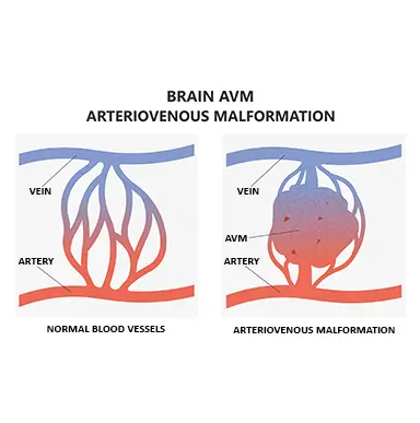
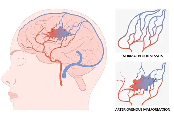
- Pediatric arteriovenous malformation (AVM) / Arteriovenous fistula (AVF): These are uncommon congenital abnormalities that cause blood to flow directly from arteries to veins bypassing the capillaries.
- Dural sinus malformation: DSM is a rare congenital malformation in children characterized by one or more dilated cranial sinuses, caused by multiple shunts between the arteries supplying the dura mater and the dural venous sinuses draining the blood from the brain.
- Infantile aneurysm: A cerebral aneurysm is a weak or thin spot on an artery in the brain that bulges out or balloons and fills with blood. Neonatal and infantile aneurysms are infrequent but can cause life-threatening intracranial bleeding if they rupture.
Rare vascular anomalies can present dramatically early during childhood, even before birth. They may cause devastating permanent neurological deficits or even life-threatening symptoms if their detection and treatment are delayed.
A multidisciplinary approach involving pediatric or obstetric, neuroradiological, neurological, and neurosurgical specialists enables prompt diagnosis and treatment of pediatric brain vascular malformations.
How Are Vascular Malformations Diagnosed?
Leading centers use cutting-edge diagnostic tools and methods that allow trained experts to diagnose brain vascular malformations accurately and instantly in children of all ages, even before they are born.
Physical examination
The physician will perform a thorough physical examination focusing on neurological and cardiological symptoms to look for signs of a brain vascular malformation.
Ultrasound
A diagnostic ultrasound, also called sonography, is a safe and painless imaging method that uses sound waves to produce images of structures within the body. A prenatal or postnatal transcranial ultrasound via the open fontanel (the skin-covered gaps on a baby's head where the skull plates meet) can diagnose cerebral anomalies, including various vascular diseases.
- Prenatal ultrasound: Pediatric specialists perform prenatal ultrasounds to diagnose Vein of Galen malformation, dural sinus malformation, and other kinds of arteriovenous malformation while the baby is still in the womb.
- Transcranial ultrasound postnatal: Pediatric specialists also conduct an ultrasound after birth to evaluate the state of vessels and form an opinion about potential brain damage or bleeding inside the brain caused by a ruptured vascular malformation.
- Cranial Doppler and Duplex ultrasound: The Doppler uses high-frequency sound waves to estimate how fast blood flows. The Duplex technique helps visualize blood flow and can confirm a diagnosis of irregular blood flow that indicates a vascular abnormality.
Computerized tomography (CT)
Computerized tomography is a series of X-ray images processed through a computer to create cross-sectional images of abnormal blood vessels in the brain and body.
- Computerized tomography (CT) scan: A computed tomography scan is most commonly used in an emergency situation to rapidly detect urgent bleeding or cerebrospinal fluid (CSF) congestion.
- Computerized tomography (CT) angiogram: If suspected that the bleeding is caused by an intracranial vascular malformation or an aneurysm, a CT angiogram can show the pathological vessels and, in some cases, the blood flow from the arteries straight to a vein (arteriovenous shunt).
Magnetic resonance imaging
Magnetic resonance imaging (MRI) combines a magnetic resonance field with computer-generated radio waves to create images of structures within your body.
- Prenatal MRI: If the ultrasound scan suspects a vascular malformation, an MRI can be used to determine a more specific diagnosis even before the baby is born. A prenatal MRI is typically performed in the 2nd or 3rd trimester of pregnancy.
- Fetal MRI: After the baby is born, an MRI is usually done prior to further therapy for vascular malformation. This helps to better localize the pathology, determine the vessels involved in the malformation, and compare the status over time.
- Magnetic resonance angiography (MRA): An MRA can create two and three-dimensional images of the neck and head vessels. Magnetic resonance angiography can accurately confirm if your child or newborn baby has any kind of vascular malformation.
Cerebral angiography
Cerebral angiography is a minimally invasive, fluoroscopic technique for visualizing blood flow in the brain's blood vessels with a contrast medium.
It is a crucial diagnostic procedure that can confirm pediatric aneurysms, dural sinus malformations, Vein of Galen malformation, cerebral arteriovenous malformations, and fistulas in the brain.
Many of these procedures require the utmost skill and care when performed on children, especially newborns. Younger children and babies have smaller blood vessels, strict radiation recommendations, and limitations on contrast medium administration which makes treatment much more challenging.
How Are Vascular Malformations Treated?
Many cerebrovascular malformations are relatively uncommon and located in highly functional brain areas. For example, the Vein of Galen malformation is located in the center of the brain, making interventional treatment very risky.
Furthermore, these birth anomalies can manifest in infants or preterm babies with inadequate body weight. Therefore, most pediatric and neurological centers prefer to wait until the infant is old enough to begin treatment. However, early treatment of children often leads to better outcomes, enhanced brain development, and ensures the survival of the newborns, particularly in severe manifestations of the Vein of Galen Malformation.
The immediate, safe, and effective treatment of vascular malformations in young infants, even hours after birth in selected cases, to relieve the child’s heart and improve cerebral circulation necessitates an exceptionally skilled and highly trained team of pediatric neuroradiologists.
Vein of Galen malformation treatment
The gold standard for treating a VOGM is embolization, a minimally invasive procedure performed under general anesthesia.
An expert interventional neuroradiologist will insert a thin, soft tube (catheter) into the arterial and venous system to advance a microcatheter to the point of the multiple arteriovenous shunts located in the central region of the brain. Then, step by step, the doctor tries to close every single feeder by placing some coils or a liquid embolic agent for blood vessel occlusion close to the point of the pathological arteriovenous connection.
To ensure treatment efficacy, interventional neuroradiologists work closely with experts in pediatrics, perinatal care, anesthesia, and neurosurgery. This multidisciplinary effort ensures that the children and newborns are continuously monitored - before, during, and after the treatment.
Pediatric brain arteriovenous malformation (AVM) / Arteriovenous fistula (AVF) treatment
Leading centers use a multidisciplinary approach involving interventional neuroradiologists, neurosurgeons, and radiation therapists to ensure the best clinical outcomes in managing AVM / AVF.
Treatment for pediatric brain AVM / AVF includes:
- Catheter embolization: This is the most common treatment option for arteriovenous fistula and arteriovenous malformation and is performed in combination with a cerebral angiogram. A microcatheter is advanced as close as possible to the fistula/malformation to occlude the pathological blood vessels supplying the AVF or AVM. The use of small coils and/or a liquid embolic agent in this procedure consequently reduces the risk of rupture.
The Pediatric Interventional Neuroradiology center at Sana Hospital Duisburg, Germany, is specialized in the super-selective microcatheterization and minimally-invasive treatment of all brain vascular malformations.
Instead of closing large parts of the vascular system, interventional neuroradiologists skillfully and precisely close the abnormal connection between the artery and the vein exactly at the point of the short circuit. Consequently, healthy vessels supplying the brain are spared, and only the abnormal vessels are closed. In some cases of AVMs, endovascular treatment combined with microsurgical resection and radiosurgery is necessary to achieve immediate care and complete recovery.
- Microsurgical resection (open vascular resection): This is an alternative treatment option for selected pediatric AVMs where the AVM’s location or the residual AVM is inaccessible to an embolization catheter. Microsurgical resection is done to separate the AVM from the neighboring tissue. The procedure utilizes a microscope to carefully clamp off the blood supply to the AVM before removing it.
- Stereotactic radiosurgery: Radiation therapists selectively perform therapeutic radiations to treat AVMs in critical brain areas or those not easily accessible with endovascular surgery or microsurgery.
Dural sinus malformation treatment
Interventional neuroradiologists follow a specific approach to occlude the shunts between the arteries supplying the dura mater, and the dilated venous sinuses, to preserve the physiological sinus drainage by:
- Inserting catheters across the artery and vein in the groin and advancing them to the level of the vessels supplying the vascular malformation.
- Positioning the microcatheters as close as possible to the arteriovenous shunts of the dural sinus malformation.
- Performing embolization of the DSM by putting some platinum coils directly in the abnormal arteriovenous connection or by applying a liquid embolic agent to achieve complete obliteration. This is done in order to retrieve the physiological blood supply to the dura and the brain and to enable the drainage of the venous system.
Infantile aneurysm treatment
The main treatment methods for brain aneurysms in children include:
- Platinum coil embolization: Coiling the aneurysm involves filling the aneurysm with tiny platinum coils that cause the blood inside the aneurysm to clot. It is done using a catheter guided along the blood vessels from the artery in the groin.
- Flow Diverter: Aneurysms can be managed by inserting an endovascular prosthesis composed of a tiny flexible mesh tube placed in the artery to cover the neck of the aneurysm. Through this, the blood flow is diverted, the regular flow is re-established, and thrombosis of the sac is determined.
- Surgical aneurysmal clipping: Surgeons place a titanium clip across the neck of the aneurysm to stop blood flow into the aneurysm.
Summary
Many newborn babies with pathological vascular malformations in the brain do not have the luxury of time and require immediate medical and surgical intervention. It is also crucial to know that a vascular malformation does not fade or disappear without treatment.
At specialized Pediatric Interventional Neuroradiology centers, like the Sana Hospital Duisburg, your child’s vascular malformation can be diagnosed precisely and treated promptly by an interdisciplinary team of specialists with expert knowledge to allow the best possible outcomes.

Dr. med. Martin Schlunz-Hendann is the Head of the Radiology and Neuroradiology department at Sana Hospital Duisburg, Germany. A certified Interventional Radiologist and Neuroradiologist with over 25 years of experience, Dr. Schlunz-Hendann leads a team covering the entire elective and emergency treatment spectrum for vascular diseases and vascular malformations of the central nervous system in adults and children. A particular focus is the interventional treatment of rare congenital cerebrovascular diseases in infants and preterm babies. As one of the largest centers in Europe for the treatment of these rare, highly complex neurovascular diseases, Dr. Schlunz-Hendann and his team treat patients from Germany and abroad.
The Pediatric Interventional Neuroradiology department at Sana Hospital Duisburg is one of the largest centers that treat very rare and highly complex brain vascular diseases in Germany and Europe. The multidisciplinary team of experts, led by chief physician Dr. Martin Schlunz-Hendann, offers a wide range of minimally invasive treatment options for infants and children of all ages with vascular malformations in the brain. Expert interventional radiologists and physicians specializing in the care of children ensure maximum safety and diagnostic accuracy. The center comprises specialists from a variety of specialties, such as neurosurgery, pediatrics, and neurology, ensuring prompt diagnosis and early treatment of children with vascular malformations in the brain.
References:
Featured Blogs

Medically Induced Coma: What It Is, How It Works, Who Benefits, and Recovery Outcomes

