Cardiac Imaging: Echo, CT, MRI, Angiography, and More

Your heart is an amazing organ. On average, it will beat more than 100,000 times each day without a break.
During your lifetime, cardiac disease might develop for a variety of reasons. Common disorders include coronary artery disease, hypertension, heart failure, cardiomyopathy, and valve disorders.
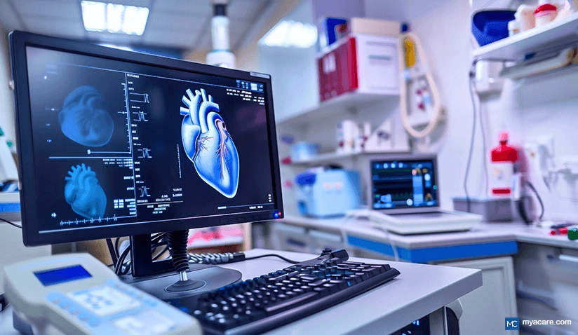
This has led to the development of many advanced imaging techniques to visualize the heart, assess its function, and search for abnormalities. Echocardiography, angiography, nuclear imaging, cardiac CT, and cardiac MRI are common practices, but how are they different? What information do they provide?
Let’s take a look at the most common heart imaging techniques used today:
Echocardiography
Echocardiography (ultrasound) is the most commonly used cardiac imaging modality around the world. It offers several advantages:
- It is cheap
- Readily available
- It is risk-free
- It can be done anywhere
- It is a rapid test
Echocardiography uses sound waves to visualize the heart, valves, great arteries, and all the structures surrounding them. Sound waves are not radioactive and are thus harmless.
There are 2 types of cardiac ultrasound:
- Transthoracic echocardiography (TTE): This is a more simple and more commonly ordered test. You will be lying down on one side, and your cardiologist will slide an ultrasound probe on your chest to see the heart from different angles on a live screen.
- Transesophageal echocardiography (TEE): This test is more complicated and is reserved for cases where a TTE is not enough. You will be sedated and a thin ultrasound probe will be inserted through your mouth and down your esophagus. Your cardiologist will move it up and down to visualize the heart on his screen.
Cardiac echography generally helps in evaluating the heart muscle, how it looks, its pumping capacity, and to see structural changes in the shape and movement of the heart. It is a critical test to diagnose heart failure and heart valve problems.
Angiography (Cardiac Catheterization)
This is another very commonly performed diagnostic cardiac test. An angiography (also called coronarography) is a more invasive test used to evaluate the coronary arteries and see if they are patent or closed.
Cardiac catheterization can be both diagnostic and therapeutic. With this intervention, your doctor can widen a blocked artery with a balloon or install a stent. Here is how a cardiac angiography goes:
- You will be sleeping on your back
- Your cardiologist will obtain access to the femoral artery (in the groin region) or radial artery (in your forearm)
- A long thin catheter will be advanced through the artery until it reaches your heart
- During this time, an X-ray machine situated above you will be taking continuous images to help guide the catheter
- Your cardiologist will occasionally inject contrast material to highlight the blood vessels and assess their patency
Cardiac Computed Tomography (Multidetector CT or MDCT)
Computed tomography (CT) scanning has been used for decades as a radiological test in most fields of medicine.
It has only recently, however, become more popular in the field of cardiology. Multidetector CT (MDCT) uses X-ray waves to assess the heart function, coronary arteries, great vessels, and heart structure.
Cardiologists use cardiac tomography to calculate the “calcium score”. By measuring the calcium in your coronary vessels, they can calculate the risk of future coronary disease and thus provide prophylactic treatment. Here’s how it goes:
- A computed tomography machine looks like a large donut.
- You will be laying on your back and will be moved through the opening for imaging.
- The test only takes a couple of minutes to complete
The beauty of multidetector cardiac CT is that it is not invasive and provides very valuable information. The only downside is radiation (although, it’s a very low dose).
Cardiac Nuclear Imaging (PET or SPECT)
Cardiac nuclear imaging is another advanced technique used to assess the structure and function of your heart. Here is how the test goes:
- First, radioactive material is injected through one of your veins
- This material will go to your heart and into your cells at different rates depending on the health of the cells and tissue.
- You will then be put into a donut machine (similar to that of the MDCT) and a series of images of your heart will be taken.
Cardiac nuclear imaging is usually combined with a stress test. This means that your heart rate will be elevated (by medication or by exercise) before the above-described procedure is done.
The test helps cardiologists assess the perfusion of the heart and the risk for coronary disease. It can also help evaluate the heart function after myocardial infarction.
Cardiac Magnetic Resonance Imaging (MRI)
Magnetic resonance is another test that has been used for decades in other medical fields.
Recently, however, magnetic resonance has become a valuable tool in the daily practice of many cardiologists. MRI is risk-free and provides very detailed information about heart function and structure.
The MRI machine is a tunnel-like machine that you will be pushed through. The machine will take many images of your heart and reconstruct them in 3-dimensions. An MRI of the heart needs 20-45 minutes to be completed.
The downside of cardiac magnetic resonance is that the machine’s tunnel is narrow and the test is loud. Many patients find this uncomfortable and might require mild sedation before the test begins.
Several cardiac MRI techniques can be performed depending on what your cardiologist wants to assess:
- Cardiac viability testing: tests the health of cardiac muscle
- Ventricular function testing: tests how good your heart is pumping blood
- Stress perfusion testing: tests the blood perfusion to your heart muscles under stress
- MRI angiography: assesses the patency and health of your coronary arteries
Depending on the type of test being done, your radiologist might or might not give you contrast material.
Conclusion
Cardiologists today have many non-invasive and invasive techniques to assess heart function. Our options are not limited anymore, thanks to the advances in medicine and technology. If you suspect that you might have heart disease or just need a check-up, discuss the benefits and risks of cardiac imaging with your cardiologist during your next visit!
To search for the best cardiology healthcare providers in Croatia, Germany, India, Malaysia, Poland, Singapore, Spain, Thailand, Turkey, the UAE, the UK and The USA, please use the Mya Care Search engine
Dr. Mersad is a medical doctor, author, and editor based in Germany. He has managed to publish several research papers early in his career. He is passionate about spreading medical knowledge. Thus, he spends a big portion of his time writing educational articles for everyone to learn.
Sources:
Featured Blogs


