
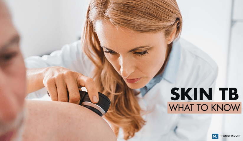
Cutaneous Tuberculosis: Recognizing the Skin Manifestations of TB
Medically Reviewed by Dr. Rae Osborn, Ph.D. - December 10, 2025
Tuberculosis (TB) is one of the oldest infectious diseases known to humanity, caused mainly by Mycobacterium tuberculosis. In the general population, tuberculosis most often involves the lungs. However, this disease can affect many other organs, including the skin.
Cutaneous tuberculosis (skin tuberculosis) is uncommon, with an estimated prevalence of 1–1.5% of TB cases involving organs other than the lungs (extrapulmonary TB). It manifests in the skin and underlying tissues. Regardless of its rarity, it is clinically significant due to diagnostic challenges, potential complications, and the ability to mimic other dermatological conditions.
Increasing awareness of this disease is vital for prevention and timely treatment, especially in parts of the world where tuberculosis is endemic.
What Is Cutaneous Tuberculosis (CTB)?
CTB refers to tuberculosis of the skin, caused by Mycobacterium tuberculosis and occasionally by Mycobacterium bovis. It develops when the bacteria infect the skin either directly or spread from another focus in the body.
Is Skin Tuberculosis Contagious?
The disease is not considered particularly infectious, as direct transmission through skin-to-skin contact is sporadically reported. The majority of infections occur through internal spread or self-inoculation in individuals already infected with tuberculosis elsewhere in the body.
What Are the Most Common Routes of Infection?
There are four common routes of cutaneous tuberculosis infection. These are:
Exogenous inoculation: This is caused by the direct entry of bacteria through cuts or wounds.
Hematogenous spread: This route involves bacteria traveling via the bloodstream from another infected site.
Contiguous spread: The infection extends from nearby lymph nodes, bones, or joints.
Endogenous: This occurs when latent bacteria reactivate within the skin itself.
What Are the Most Important Risk Factors?
The common risk factors for skin TB infection are:
- Immunocompromised states (HIV, organ transplantation, long-term corticosteroid use)
- Malnutrition
- Poor living conditions and close exposure to active TB cases
- Extremes of age and, in some cases, female gender
Types of Cutaneous Tuberculosis
The cutaneous tuberculosis classification includes several different forms. Each of these has distinctive clinical features:
Primary Inoculation Tuberculosis
This type is also called primary cutaneous tuberculosis. It develops when the bacteria directly enter the skin of a person without prior exposure to tuberculosis. The type commonly presents as a chancre with regional lymph node involvement.
Lupus Vulgaris
The most common form of tuberculosis skin disease manifests as slowly progressing reddish-brown plaques (also known as apple-jelly nodules). These are most often located on the face and extremities.
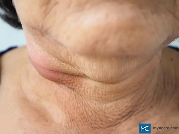
Scrofuloderma
Scrofuloderma is caused by the direct extension of tuberculosis infection from underlying lymph nodes, bones, or joints to the skin. A typical presentation includes nodules that soften into ulcers with discharge.
Tuberculosis Verrucosa Cutis
This type of skin TB is most commonly seen in previously sensitized individuals. These are usually healthcare workers or farmers. The infection manifests as warty skin lesions, mostly affecting the hands, lower legs, or feet.
Orificial Tuberculosis
Orificial TB is a rare but severe type of cutaneous tuberculosis that affects mucocutaneous junctions such as the mouth, anus, or genitalia. The type is usually seen in patients with advanced internal TB.
Acute Miliary Tuberculosis of the Skin
This type is also known as cutaneous miliary tuberculosis. It is caused by widespread blood-borne dissemination of TB. The infection produces tiny plaques or ulcers over large areas of the skin.
Other (Rare) Forms of Cutaneous Tuberculosis
There are also some very rare forms of cutaneous TB. These include:
- Papulonecrotic tuberculid or hypersensitivity reaction leading to necrotic papules.
- Lichen scrofulosorum manifests as grouped papules. Usually, in children or adolescents with underlying TB.
- Erythema Induratum of Bazin (EIB) is associated with TB hypersensitivity. It presents as nodules, usually on the legs.
Clinical Features of Cutaneous TB
The presentation of skin tuberculosis varies with type, but common patterns include:
Skin Lesions
Skin lesions may include nodules, plaques, ulcers, or warty growths.
Typically Affected Sites
The face, neck, extremities, and lymph node regions are commonly affected.
Common Skin Tuberculosis Symptoms
These typically include pain, swelling, discharge, scarring, and pigmentation changes.
Systemic Signs
Systemic signs include fever, weight loss, night sweats, and cough (if there is active TB elsewhere).
Diagnosis of Cutaneous Tuberculosis
Diagnosing tuberculosis of the skin requires a combination of clinical and laboratory evaluation, which typically involves:
Clinical Examination
This usually involves a physical check-up and taking the patient’s medical history, noting all the risk factors and potential exposure to tuberculosis infection.
Laboratory Tests
This may include a skin biopsy to detect granulomas, culture (or PCR) to confirm the presence of Mycobacterium tuberculosis, and Ziehl-Neelsen staining to demonstrate acid-fast bacilli.
Imaging
This can be useful if bones or deeper tissues are affected.
Differential Diagnosis
It is absolutely essential, for accurate diagnosis and effective management, to differentiate cutaneous tuberculosis (CTB) from other dermatological conditions with overlapping clinical features. Here’s an overview of infectious and noninfectious diseases that can mimic CTB:
Leprosy
Both CTB and leprosy may present with chronic skin lesions and nerve involvement. However, leprosy typically causes anesthetic patches with loss of sensation and peripheral nerve thickening, while CTB lesions are usually ulcerative and associated with underlying lymphadenopathy.
Deep Fungal Infections
Conditions such as sporotrichosis, chromoblastomycosis, and blastomycosis can resemble CTB with nodules, ulcers, or verrucous plaques. Fungal infections are distinguished by the identification of fungal elements on microscopy or culture, and they often respond to antifungal therapy rather than anti-tubercular treatment.
Sarcoidosis
Both sarcoidosis and CTB may present with granulomatous lesions. However, sarcoidosis granulomas typically lack necrosis.
Psoriasis
Chronic plaque psoriasis can mimic lupus vulgaris (a form of CTB) due to scaly plaques, but psoriasis lesions are usually well-demarcated, silvery, and symmetrically distributed on extensor surfaces. Histopathology and the absence of granulomas help distinguish psoriasis from CTB.
Leishmaniasis
Cutaneous leishmaniasis can present as chronic ulcers or nodules similar to CTB. Travel or residence in endemic regions, demonstration of Leishmania amastigotes on microscopy, and response to antileishmanial therapy confirm the diagnosis.
Lupus Erythematosus
Discoid lupus erythematosus may mimic lupus vulgaris, with erythematous plaques and scarring. However, lupus erythematosus lesions show follicular plugging, interface dermatitis, and positive lupus-specific serology (e.g., ANA).
Atypical Mycobacterial Infections
Non-tuberculous mycobacteria (such as Mycobacterium marinum or M. ulcerans) can cause skin infections resembling CTB. Differentiation relies on microbiological culture or PCR for molecular identification, as well as the patient’s history of exposure to contaminated water or trauma.
What Are the Treatment Options?
The treatment of cutaneous tuberculosis does not differ much from that of systemic tuberculosis. Primarily, it is based on multi-drug anti-tubercular therapy (ATT). The main goal is to eradicate Mycobacterium tuberculosis. Additionally, it is essential to prevent relapse and restore healthy skin function and appearance.
What Is Standard Anti-Tubercular Therapy (ATT)?
Both the shorter and conventional regimens fall into the standard treatment approach category, according to the WHO’s consolidated guidelines from 2024[18], depending on patient eligibility.
The four-month regimen is preferred where appropriate, especially for adolescents and adults (older than twelve) with confirmed drug-susceptible pulmonary tuberculosis and no evidence of resistance or severe disease. The regimen includes two phases:
- Intensive phase (two months): Isoniazid (H), Rifapentine (P), Pyrazinamide (Z), and Moxifloxacin (M).
- Continuation phase (two months): Isoniazid, Rifapentine, and Moxifloxacin.
The conventional six-month regimen is suitable for patients who are not eligible for the shorter regimen. These can be individuals with extrapulmonary TB, cavitary disease, or suspected resistance. In such cases, there are also two distinct phases of treatment:
- Intensive phase (two months): Isoniazid (H), Rifampicin (R), Pyrazinamide (Z), and Ethambutol (E).
- Continuation phase (four months): Isoniazid and Rifampicin.
Treatment duration may be extended in specific cases, such as relapse, treatment interruption, or severe disease.
Regular monitoring to assess the therapeutic response is crucial, as there can be side effects.
Clinical and laboratory monitoring are also important for evaluating the healing of skin tuberculosis symptoms and checking liver function.
Management of Complicated or Refractory Cases
The standard treatment may not be effective when drug-resistant strains of bacteria are present. Such cases create the need for second-line anti-tubercular drugs. This also calls for specialist supervision.
Second-line treatment options have evolved and developed over time. Before, the protocols often included fluoroquinolone antibiotics (e.g., levofloxacin, moxifloxacin) or injectable medications, such as amikacin and capreomycin. The modern approach, however, has replaced these with shorter and fully oral treatments which are safer and more effective, as recommended by the WHO.
The latest WHO guidance emphasizes all-oral combination regimens based on bedaquiline, pretomanid, linezolid, and moxifloxacin (when bacteria are still sensitive to it). In cases where fluoroquinolones are no longer effective, treatment can still be delivered orally by using bedaquiline, pretomanid, and linezolid alone.
For patients who cannot use the shortest regimen, a slightly longer nine-month oral course may be recommended. This version typically combines bedaquiline with a fluoroquinolone and additional oral agents such as clofazimine, high-dose isoniazid, ethambutol, and pyrazinamide (depending on individual drug-sensitivity results and clinical judgement).
Surgical Intervention
Surgery plays a supportive role in certain cases of cutaneous tuberculosis and might involve:
- Drainage or debridement of abscesses and sinus tracts
- Excision of necrotic tissue or non-healing ulcers
- Reconstructive surgery or skin grafting for cosmetic or functional restoration (skin lesions in tuberculosis can leave deep scars)
Surgical procedures can only follow ATT to minimize the bacterial burden and prevent recurrence.
Potential Complications and Prognosis
With appropriate therapy, prognosis is generally favorable, and patients often recover well. However, untreated or advanced symptoms may lead to:
- Permanent scarring and pigmentation changes
- Pregnancy complications (rare, but careful monitoring is required)
- Malignant transformation into squamous cell carcinoma or co-occurrence of CTB with lymphoma (rare but reported)
- Progression to systemic or disseminated TB in immunocompromised patients
Prevention and Awareness
Prevention strategies for tuberculosis skin disease overlap with general TB control:
- Early detection and treatment of systemic TB to prevent spreading
- Maintaining hygiene and proper wound care to reduce the chances of inoculation
- Raising awareness in high-risk and endemic areas
- Strengthening nutrition and immunity through community health measures
- Bacille-Calmette-Guérin vaccination in countries where TB is prevalent
The Bottom Line
Cutaneous tuberculosis is a rare but significant manifestation of tuberculosis affecting the skin. The symptoms can resemble those of many other skin disorders. Therefore, early recognition and proper diagnosis are crucial for effective management. With timely treatment initiation, most individuals respond well to standard anti-tubercular therapy and heal completely.
To search for the best Dermatology Healthcare Providers in Croatia, Germany, Greece, India, Malaysia, Singapore, Slovakia, Spain, Thailand, Turkey, the UAE, UK, the USA, please use the Mya Care search engine.

Dr. Rosmy Barrios is an aesthetic medicine specialist with international work experience. She earned her physician diploma at the Universidad Del Norte’s School of Medicine in Barranquilla, Colombia, and her specialty at John F. Kennedy University in Buenos Aires, Argentina. Dr. Barrios is a member of the Pan-American Aesthetic Medicine Association (PASAM) and the Union Internationale de Médecine Esthétique (UIME). She is an expert health writer with keen interests in aesthetic medicine, regenerative aesthetics, anti-aging, fitness, and nutrition. Currently, Dr. Barrios heads the Regenerative Aesthetics department at a renowned Internal Medicine clinic based in Belgrade, Serbia.

Dr. Rae Osborn has a Ph.D. in Biology from the University of Texas at Arlington. She was a tenured Associate Professor of Biology at Northwestern State University, where she taught many courses to Pre-nursing and Pre-medical students. She has written extensively on medical conditions and healthy lifestyle topics, including nutrition. She is from South Africa but lived and taught in the United States for 18 years.
References:
Featured Blogs

Medically Induced Coma: What It Is, How It Works, Who Benefits, and Recovery Outcomes

