
What Is Granuloma Annulare? Causes, Diagnosis & Treatment Options
Medically Reviewed by Dr. Asif Baliyan, MD - January 30, 2026
Granuloma annulare is a benign but chronic skin condition characterized by a circular skin rash in the form of small and firm bumps. Hence the name “annulare”, the Latin word for ring. The condition involves inflammation in the dermis due to a localized breakdown of collagen triggered by the immune system. Experts still cannot precisely define granuloma annulare. However, it is thought to be a hypersensitivity reaction involving immune dysregulation
The condition is relatively uncommon but tends to affect individuals of all ages and both genders, with a slightly higher prevalence in women. Granuloma annulare can occur in childhood, particularly in its localized form, although it is relatively uncommon overall; most cases present before age 30. While it often manifests as a single, small ring on the hands or feet, it may also occur in a widespread form called disseminated granuloma annulare, affecting large portions of the body.
Granuloma annulare is not officially classified as an autoimmune disease. However, it shares characteristics of an immune-mediated inflammatory reaction. Some studies link granuloma annulare to diabetes and other autoimmune or metabolic disorders such as thyroid disease and rheumatoid arthritis. All of this further suggests that the immune system could play a significant role in the disease’s development and recurrence.
What Are the Signs and Symptoms of Granuloma Annulare?
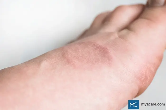
Lesions usually start as small papules that expand outward to form smooth, firm rings. Patients often describe them as bumps in a circle on the skin or a circular skin rash. Granuloma annulare on the hands, elbows, knees, ankles, or feet is more common. Facial involvement and scalp lesions are observed less often, but are possible.
Most patients experience no pain, though a few describe the rashes as being itchy. The lesions are usually skin-colored, pink, or red, but may appear darker or purplish, especially with granuloma annulare on dark skin.
Types of Granuloma Annulare
The condition can appear in several distinct clinical patterns, each with unique features and common locations:
Localized Granuloma Annulare
This is the most frequent form, as it accounts for two-thirds of all cases and represents approximately 75% of the reported cases of granuloma annulare. The condition presents as small, smooth papules arranged in a circular or arc-like pattern. These are often described as bumps in a circle on the arm or on the back of the hands and feet. The center of the ring may look slightly depressed or normal in color. Localized GA is usually asymptomatic and self-limiting. It often resolves within 6 to 24 months.
Generalized (Disseminated) Granuloma Annulare
In this type, multiple lesions appear across the trunk, neck, or extremities. These may merge to form larger patches or rings. This form is more common in adults and may persist for several years. Biopsy confirmation is often required because disseminated granuloma annulare can resemble other chronic dermatoses.
Subcutaneous or Deep Granuloma Annulare
The condition can present as firm and painless nodules under the skin, i.e., subcutaneously. Deep granuloma annulare, as it is also known, is most often seen in children, especially on the shins, scalp, hands, or buttocks. The overlying skin appears normal or slightly elevated. These nodules are benign but can worry parents because they feel like small lumps.
Perforating Granuloma Annulare
A rarer subtype, perforating granuloma annulare, involves small papules that develop a central plug. Over time, the skin’s surface breaks down, allowing material from deeper layers to “perforate” outward. It often affects the hands and forearms and can leave small scars after healing.
Patch Granuloma Annulare
This variant presents as smooth, flat, or slightly scaly reddish-brown patches. Because it lacks the typical raised ring, it may be confused with eczema or nummular dermatitis. The lesions are often seen on the trunk and thighs and may cause mild itching.
Interstitial Granuloma Annulare
Under the microscope, this form shows a diffuse inflammatory pattern, hence the name interstitial granuloma annulare. Clinically, it can appear similar to the patch or localized types but may persist longer and respond differently to treatment.
Atypical Granuloma Annulare
In some individuals, the condition manifests as non-ring patterns or as thick plaques, linear lesions, or even ulcerative forms. These atypical presentations are uncommon and can occur in immunocompromised or diabetic patients.
Causes and Risk Factors
The condition often begins after minor skin trauma, insect bites, or infections. Inflammation causes the skin’s collagen to degrade, forming small clusters of immune cells that create the raised circular lesions.
What Are the Known Triggers?
Common triggers include:
- Emotional or physical stress, which can activate immune responses and provoke outbreaks
- Sun exposure or minor skin injuries
- Hormonal fluctuations
- Vitamin D deficiency or imbalance
Systemic Associations
Granuloma annulare may accompany systemic conditions such as:
- Diabetes mellitus
- Thyroid disease (especially hypothyroidism)
- Dyslipidemia or metabolic syndrome
Granuloma Annulare and Cancer
In some patients, especially older adults, disseminated granuloma annulare has been observed to develop as a paraneoplastic reaction. That means the skin eruption appears as an immune response to an underlying malignancy.
Reported associated cancers include:
- Lymphomas (especially Hodgkin’s and non-Hodgkin’s)
- Leukemia
- Breast cancer
- Prostate cancer
- Lung cancer
- Gastrointestinal cancers
In such cases, the skin lesions may improve or disappear once the cancer is treated. This further supports the idea of an immune-mediated connection rather than a direct cause.
Infectious and Genetic Factors
The following are a few commonly asked questions about the infectious and genetic factors that may contribute to granuloma annulare:
What viral infections cause granuloma annulare?
Case reports link it to Epstein–Barr virus, hepatitis B and C, HIV, and occasionally after vaccinations.
Can parasites cause granuloma annulare?
This is extremely uncommon, but chronic parasitic infections may theoretically trigger immune responses.
Is granuloma annulare hereditary?
It usually occurs sporadically, though familial clustering has been noted in rare cases.
Is granuloma annulare related to lupus?
No. While the rashes can look similar, lupus involves systemic autoimmunity, whereas granuloma annulare is confined to the skin.
Are there any possible complications?
The condition is benign, not contagious, and rarely leaves permanent damage. However, after healing, some people develop residual hyperpigmentation or lighter patches, particularly those with dark skin.
How Is Granuloma Annulare Diagnosed?
Diagnosis is usually straightforward based on appearance (smooth, firm, ring-shaped papules without scaling). A biopsy can confirm granuloma annulare and rule out mimics such as fungal infections or autoimmune skin conditions.
Differential Diagnosis
Granuloma Annulare vs Ringworm (Tinea Corporis)
Tinea corporis may have the closest resemblance to granuloma annulare. It is a superficial dermatophyte infection that also presents with annular lesions. However, ringworm typically has a scaly, raised, and active border with central clearing. Also, the lesions often itch and may spread centrifugally. A potassium hydroxide (KOH) preparation from skin scrapings helps differential diagnosis as it will reveal fungal hyphae. Ringworm lesions respond rapidly to topical or systemic antifungal therapy.
Granuloma Annulare vs Psoriasis
Unlike granuloma annulare, where scaling is absent, psoriasis usually presents with well-demarcated erythematous (abnormally reddened) plaques covered by thick, silvery scale. These commonly appear on extensor surfaces, scalp, and sacral area, and are usually not ring-shaped. Psoriasis is also often associated with nail changes (pitting or onycholysis) and may cause itchiness.
Nummular Eczema vs Granuloma Annulare
Nummular eczema produces coin-shaped, erythematous, scaly, and intensely itchy plaques. These are often oozing or crusting in acute stages, unlike GA lesions, which are typically asymptomatic, firm, and non-scaly. Xerosis (dry skin), irritants, or atopy (an inherited tendency to develop allergies) commonly trigger the nummular eczema flare-ups.
Granuloma Annulare vs Cutaneous Lupus Erythematosus
Cutaneous lupus may produce annular or discoid lesions, similar to GA. However, this is often accompanied by photosensitivity, dyspigmentation, scarring, ulceration, or systemic symptoms, such as fatigue or arthralgia (joint pain). Additionally, serologic abnormalities (ANA, anti-dsDNA) and involvement of other organs may be present. Histopathology and immunofluorescence help distinguish lupus from granuloma annulare.
Actinic Granuloma vs Granuloma Annulare
Actinic granuloma is strongly associated with sun (UV) exposure. It usually presents as annular plaques with raised borders and central atrophy, in sun-exposed areas of older individuals. Actinic granuloma is thought to be related to solar elastosis, and it shows elastophagocytosis on histology.
Granuloma Annulare vs Sarcoidosis
Cutaneous sarcoidosis can mimic granuloma annulare with papules and plaques, but lesions are often reddish-brown or violaceous (violet-purple in color). They can also be accompanied by systemic involvement, affecting the lungs, lymph nodes, or eyes. Biopsy helps differential diagnosis as it shows non-caseating granulomas without the palisading pattern typical of granuloma annulare.
Granuloma Annulare vs Erythema Annulare Centrifugum
Erythema annulare centrifugum is a reactive annular eruption that features expanding rings with a trailing scale on the inner border. However, on visual inspection, they appear different from the granuloma annulare-associated smooth, non-scaly lesion surface. Erythema annulare centrifugum is commonly associated with infections, medications, or internal disease.
Granuloma Annulare vs Necrobiosis Lipoidica
Necrobiosis lipoidica presents with yellow-brown atrophic plaques with telangiectasias. It usually affects the shins and is strongly associated with diabetes mellitus. Lesions may ulcerate and have a shiny, thinned appearance, unlike granuloma annulare, which remains firm and intact.
What Are the Treatment Options?
Many cases improve spontaneously within months to two years. Observation is often recommended for mild, asymptomatic, localized lesions, particularly in children.
Topical and Local Therapies
- Topical corticosteroids can reduce inflammation and help flatten lesions. Applied under occlusion, they are the first-line therapy.
- Calcineurin inhibitors (e.g., tacrolimus or pimecrolimus) are useful alternatives for delicate areas like the face, granuloma annulare on the finger, or intertriginous regions.
- Intralesional corticosteroid injections are beneficial for small, stubborn plaques on the hands or feet.
Systemic and Phototherapy Treatments
With disseminated granuloma annulare or widespread lesions that are causing discomfort or cosmetic concern, stronger treatments may be required:
- Oral medications, such as hydroxychloroquine, isotretinoin, dapsone, or short courses of oral corticosteroids, can help control inflammation.
- Phototherapy with narrowband UVB or PUVA therapy may induce remission in generalized cases.
- Systemic immunomodulators like Methotrexate or cyclosporine are used in resistant disease, particularly in adults with autoimmune comorbidities.
Procedural and Cosmetic Options
- Cryotherapy (freezing) may be effective for isolated lesions but may also cause hypopigmentation.
- Laser ablation using fractional and pulsed-dye lasers helps remodel the skin and reduce residual discoloration.
Emerging and Experimental Treatments
Recent advances in dermatology have produced new treatments for granuloma annulare, with a focus on immune modulation:
- JAK inhibitors (tofacitinib, ruxolitinib)
Early studies show significant improvement in chronic, treatment-resistant cases.
- Low-dose naltrexone
Promotes immune regulation and may reduce inflammation.
- Biologic therapies
Agents targeting TNF-alpha (adalimumab, etanercept) have shown promise for severe or recurrent granuloma annulare.
Diet and Home Remedies
Although diet alone does not cure granuloma annulare, certain changes may support healing and reduce recurrence. Here are some suggestions:
- Limiting alcohol, caffeine, and processed foods
- Following an anti-inflammatory or gluten-free diet
- Ensuring adequate vitamin D and antioxidant intake
- Using gentle home remedies, such as aloe vera, turmeric, or green tea extracts, to soothe the skin
Living With Granuloma Annulare
While granuloma annulare may persist or recur, it remains a non-contagious and benign condition. Therefore, identifying and addressing potential triggers, such as stress, infections, or underlying systemic diseases, can reduce recurrences.
Patients with granuloma annulare on the hands or with facial involvement may experience cosmetic distress. However, reassurance is vital as the condition often resolves naturally or responds well to treatment.
In children, subcutaneous granuloma annulare nodules typically disappear completely without therapy. In adults, periodic flare-ups can occur but are manageable with proper care.
The Bottom Line
Although the cause of granuloma annulare remains uncertain, it is associated with immune activity, stress, and metabolic factors, such as diabetes. Most cases resolve spontaneously, but persistent or widespread forms may benefit from topical or systemic treatment.
To search for the best Dermatology Healthcare Providers in Croatia, Germany, Greece, India, Malaysia, Singapore, Slovakia, Spain, Thailand, Turkey, the UAE, UK, the USA, please use the Mya Care search engine.

Dr. Rosmy Barrios is an aesthetic medicine specialist with international work experience. She earned her physician diploma at the Universidad Del Norte’s School of Medicine in Barranquilla, Colombia, and her specialty at John F. Kennedy University in Buenos Aires, Argentina. Dr. Barrios is a member of the Pan-American Aesthetic Medicine Association (PASAM) and the Union Internationale de Médecine Esthétique (UIME). She is an expert health writer with keen interests in aesthetic medicine, regenerative aesthetics, anti-aging, fitness, and nutrition. Currently, Dr. Barrios heads the Regenerative Aesthetics department at a renowned Internal Medicine clinic based in Belgrade, Serbia.

Dr. Asif Baliyan is a doctor and clinical researcher with over a decade of experience in evidence-based diagnostic medicine. A Consultant at a tertiary care hospital in New Delhi, he also serves as a medical reviewer, ensuring healthcare content remains accurate, ethical, and aligned with current clinical guidelines.
References:
Featured Blogs

Medically Induced Coma: What It Is, How It Works, Who Benefits, and Recovery Outcomes

