
Excretory System Overview: How the Body Removes Waste

Originally Medically Reviewed by Dr. Sony Sherpa (MBBS) - August 13, 2024
Fact Checked and Updated by Dr. Dora Matis - March 18, 2026
The excretory system removes metabolic waste and maintains fluid, electrolyte, and acid–base balance essential for survival. The urinary (renal) system plays a central role through kidney filtration and bladder storage, while also regulating hormones and blood pressure. Additional excretory functions are carried out by the digestive system, lungs, and skin through feces, exhalation, and sweat.
Introduction
The urinary system, the main component of the excretory system, is essential for survival, as it maintains internal homeostasis through the elimination of metabolic waste products and the regulation of fluid and electrolyte balance. This system is primarily involved in the removal of waste products from the body. Without adequate renal function, metabolic waste products accumulate, leading to life-threatening complications.
The following overview discusses the excretory system, with the main emphasis on the urinary system.
Elimination vs. Excretion
Excretion is the process by which the body removes waste products produced during normal cellular metabolism and is essential for maintaining balance within the body.
Cells take in nutrients and use them to produce functional products in response to cellular signals. Signals consist of a wide array of compounds, including hormones, microbial by-products, and certain nutrients (such as minerals, vitamins, amino acids, and other peptides) that act as regulatory signals. Additionally, nerve impulses and environmental cues such as temperature and pH affect certain cell cultures in ways that generate signals in response. Signaling compounds instruct the cell what products to produce from the building blocks it receives. Some of these signals ultimately affect the cell’s DNA by turning specific genes on or off.
Because waste is produced by cells throughout the body, many organs contribute in some way to removing it. Thus, the term elimination is used to describe cellular excretion – the waste products are typically eliminated from the cell and moved along to an organ that can process them.
Components of the Excretory System
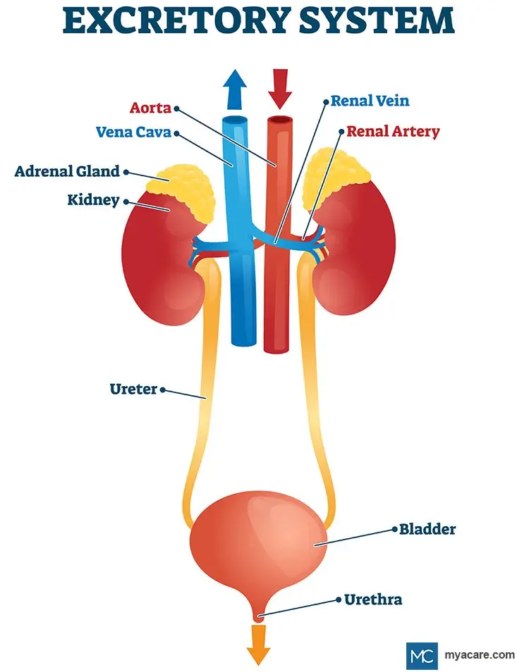
The main organs that process cellular waste products for excretion (complete removal from the body) are the kidneys, the lungs, and also the digestive tract (mainly the colon with its gut microbiome), and the skin. The kidneys produce urine by filtering waste products from the blood, and the bladder stores this urine until it is expelled from the body; together, these organs are part of the urinary system.
The urinary system is perceived as being the most important part of the excretory system as a whole and is discussed below. The other systems involved in excretion are also reviewed.
The Urinary System
The urinary system is responsible for processing waste products from the blood, filtering them in the kidneys, and excreting them via the ureter (tube connecting the kidneys to the bladder), bladder, and urethra. Another name for the urinary system is the renal system.
Aside from the filtration and elimination of blood waste products, the renal system helps to maintain healthy blood pressure, blood pH, electrolyte balance, and fluid balance.
Kidneys
The kidneys take the majority of cellular waste from the body, transported to them via the bloodstream. They produce urine, which travels to the bladder where it is stored until it is expelled from the body.
The kidneys receive a large blood supply through the renal arteries, which branch from the abdominal aorta. They require optimal oxygenation for energy production in order to maintain optimal homeostasis in the body.
Regulation of Kidney Function
The kidneys are regulated by hormones, nerve impulses, as well as their own internal regulatory mechanisms.
1. Endocrine and Nervous System Regulation
The kidneys contain neuroendocrine cells that communicate with the brain, sending signals that influence kidney function and responding to signals from the brain.
The hypothalamus in the brain receives a wide variety of signals relevant to kidney function by monitoring nerve impulses and blood composition. In response to these stimuli, it sends signals to the pituitary, which then, in turn, releases hormones that the kidney responds to by modulating its functions.
Sympathetic nerve impulses cause smooth muscles in the kidney’s arterioles to relax or contract, helping adjust blood flow and filtration according to the body’s needs.
2. Auto-Regulation
In the absence of neuro-endocrine involvement, the kidneys are still able to function and appear to have their own form of auto-regulation.
Auto-regulation is a homeostatic mechanism that is usually referred to in the context of the cardiovascular system, in which blood vessels relax or contract as a result of increased or decreased blood pressure, respectively. When applied to the kidneys, auto-regulation of kidney blood vessels alters the rate at which they eliminate and reabsorb waste products, thereby regulating kidney function.
Kidney Function
The two main functions of the renal system are elimination and osmoregulation, which each simultaneously serve a variety of other functions when fulfilled by the kidneys.
1. Elimination
Elimination refers to the removal of waste products. In terms of kidney function, elimination refers to their removal from the bloodstream. The kidneys filter the blood, producing urine, which is collected in the bladder for excretion. The kidneys filter approximately up to 200 L or 53 gallons of water from the blood every day.
A significant portion of bodily waste is derived from the liver. The liver is involved in many processes pertaining to bio-transformation, in which nutrients or cellular metabolites are transformed into further useful products and waste products. Urea is synthesized primarily in the liver before it makes its way via the bloodstream to the kidneys and to a lesser extent, the skin.
The kidneys filter blood plasma, removing urea along with excess water, electrolytes (such as sodium, potassium, calcium, magnesium, and chloride), and other waste products. Ammonia, uric acid, creatinine, pigments, and inorganic salts also form part of urine.
The main active component of the kidney would be the nephrons, which are responsible for reabsorbing useful substances and eliminating unnecessary ones. Nephrons consist of intricate renal tubes, which are designed to recycle metabolites in the blood and excrete the rest. Anything that can be recycled is typically sent back to the bloodstream, while the rest combines with water to produce urine. This is achieved through selective membrane permeability. Mineralocorticoids and certain other substances alter the permeability of the membranes inside nephrons.
Different portions of each nephron are involved in different aspects of elimination. Nephrons are anatomically divided into the following sections:
- The Glomerulus is a small tuft of capillaries inside the renal corpuscle of each nephron. It filters blood plasma, allowing water, ions, and small molecules, including wastes, to enter the nephron while keeping blood cells and proteins in the bloodstream.
- Renal Corpuscles are tiny, yet bulbous structures that surround the capillary loops of the glomerulus. The part surrounding the glomerulus is called the glomerular capsule or Bowman’s capsule. Blood is allowed to fill the spaces around the glomerulus. A thin membrane separates this blood from urea and other waste products that move through to the internal compartment of the renal corpuscle, referred to as Bowman’s Space. Once blood plasma is filtered, it passes back out of the kidneys and into circulation. Unfiltered blood enters renal corpuscles through the afferent arterioles and filtered blood leaves through the efferent arterioles.
- Renal Tubules consist of the proximal convoluted tubule, the loop of Henle, and the distal convoluted tubule, followed by the collecting duct. These portions of the nephron further refine elimination and aid in osmoregulation. As the fluid passes through these tubules, the lining cells help to recover useful substances, such as water, minerals, and nutrients, returning them to the bloodstream. The efferent arteriole gives rise to a capillary network apposed to the renal tubules, facilitating the exchange of metabolites, minerals, and water.
- The Proximal Convoluted Tubule is the first part of the renal tubule. It reabsorbs water, glucose, amino acids, electrolytes, and other useful compounds back into the blood. It also actively secretes substances such as hydrogen ions, creatinine, penicillin, and other organic molecules, helping maintain blood pH and remove wastes.
- The Loop of Henle descends from the proximal tubule and then curves back upward to connect to the distal tubule. The thin descending limb is permeable to water, allowing water to leave into the surrounding tissue. The thick ascending limb is impermeable to water but actively reabsorbs sodium, potassium, and chloride, helping the kidney concentrate urine and maintain electrolyte balance.
- The Distal Convoluted Tubule fine-tunes the filtrate by reabsorbing sodium and regulating electrolyte balance. It also secretes hydrogen ions and potassium to help maintain acid-base balance. Water reabsorption in the later segments depends on antidiuretic hormone (ADH). After further adjustment in the collecting duct, the remaining fluid becomes urine.
- Collecting Ducts technically do not form part of nephrons, but are nonetheless continuous with them. They eliminate urine from the nephron by emptying it into the ureter for transportation to the bladder. They also play an important role in the final adjustment of water and electrolyte balance.
There are two types of nephrons: cortical and juxtamedullary. Cortical nephrons make up about 85% of all nephrons and are located mainly in the outer cortex. Juxtamedullary nephrons are located near the corticomedullary junction and only comprise up to 15% of the nephrons inside the kidney, and have long loops of Henle that extend deep into the medulla. Each nephron functions independently.
2. Hormone Production
The kidneys have important endocrine functions and produce hormones that regulate blood pressure, osmosis, electrolyte balance, and more. These hormones include:
- Renin, which is required for osmoregulation, forming part of the renin-angiotensin system.
- Erythropoietin Hormone, which stimulates the growth and development of red blood cells from stem cells in the bone marrow.
- Vitamin D3 (cholecalciferol), which is a prohormone that is converted in the body to its active form, 1,25-dihydroxyvitamin D (calcitriol). Calcitriol acts as a hormone and exerts effects throughout the body. Calcium homeostasis, the immune system, growth and development, as well as cellular energy production, are regulated in part by the actions of vitamin D3. Vitamin D3 is synthesized in the skin during exposure to ultraviolet B radiation and can also be obtained from dietary sources.
3. Osmoregulation
The kidneys regulate body fluid balance by controlling the reabsorption and excretion of electrolytes and water.
Mineral ions govern the movement of fluids in and out of cells, being the master coordinators of osmosis in the body. Thus, disturbances in the balance of cellular (internal) and blood (external) mineral concentrations determine how much water the kidneys withhold to maintain osmoregularity. Sodium is the principal determinant of extracellular fluid volume and water retention, while potassium primarily regulates intracellular fluid balance. Water levels are also involved in conserving blood volume, a factor that is intimately linked with cardiovascular function and blood pressure.
Osmoregulation is further governed by a group of hormones produced at various sites of the body. These hormones include:
- Renin is produced in the kidneys and is required by the renin-angiotensin system to regulate osmosis, blood pressure, and blood volume. Renin chemically breaks down angiotensinogen, a substance that is primarily produced by the liver to create angiotensin. Other organs can also produce angiotensinogen, suggesting that the rest of the body may also be involved in regulating kidney function and blood pressure. It is also possible that this precursor is involved in blood vessel growth, yet evidence is inconclusive. Renin is triggered in response to lowered renal blood pressure or low sodium levels in the renal tubules.
- Angiotensin I and II are created from the interactions of renin and angiotensinogen, a protein made in the liver. Renin transforms angiotensinogen into angiotensin I, which is then further converted into angiotensin II by Angiotensin- Converting Enzyme. Angiotensin II constricts blood vessels in order to maintain optimal blood pressure. When levels of angiotensin II are high, renin production is low, and vice versa. Angiotensin II helps the kidneys maintain filtration by constricting the small vessels leaving the glomerulus and increases sodium reabsorption through aldosterone. Elsewhere in the body, it constricts blood vessels, which raises blood pressure. This hormone also binds and acts in the hypothalamus to increase the sensation of thirst, as well as water consumption.
- Aldosterone is an adrenal hormone that is released in response to angiotensin II. It works to increase sodium reabsorption by modulating the mineral balance inside the kidneys. Aldosterone is known as a mineralocorticoid in this respect.
- Antidiuretic Hormone is released from the pituitary in response to hypothalamic osmotic feedback, subsequent signaling, and blood levels of angiotensin II. This hormone acts on the nephrons of the kidneys, instructing them to increase permeability in the collecting ducts and to reabsorb more water. This increases bodily water retention and concentrates or even withholds urine.
Other hormones can also affect kidney function, such as endothelin (a vascular protein) and norepinephrine, which promote vasoconstriction and a decrease in the filtration rate. Vasodilators such as nitric oxide, histamine, as well as dopamine acting on renal dopaminergic receptors, can increase renal blood flow, which tends to increase or preserve glomerular filtration rate. Inflammatory cytokines also influence renal vascular tone depending on context.
Bladder
The bladder is an organ that takes urine from the kidneys via the ureter, storing it until urination is possible. The thick membrane of the bladder comprises three layers of muscle tissue known as the detrusor muscle, which facilitates its functions. The bladder has two sphincters at its outlet: the internal sphincter and the external sphincter. Together, they control the flow of urine into the urethra.
Bladder Function
The bladder serves two main functions, with a potential third function:
1. Storage
When the bladder is not in use, the detrusor muscle remains relaxed while the sphincters remain contracted. This ensures that urine remains inside the bladder, storing it until excretion takes place. Storage of urine is particularly important during the night when we’re asleep in order to promote better rest and rejuvenation.
Bladder volume and pressure are controlled through the nervous system via the actions of neuroendocrine cells, neurons, and nerve impulses. When pressure in the bladder rises due to urinary retention, nerves connected to the bladder relay sensory information to the brain via the pelvic girdle and spinal cord. Through this feedback, we become aware that we need to urinate. Other nerve fibers respond to other signals, such as chemical irritation, similarly alerting the brain, which then acts to mediate bladder function appropriately.
2. Excretion
During urination, the sphincters relax and open, while the bladder contracts alongside relevant portions of the urethra, facilitating urinary excretion. The contraction of the bladder also pinches closed the opening to the ureters, ensuring that the kidneys don’t experience urinary reflux during urinary excretion.
Aside from being governed via central nervous system feedback, urination is also governed by the peripheral nervous system. The sympathetic nervous system prevents urination from occurring, and the parasympathetic nervous system encourages it. In this respect, feeling stressed can cause urinary retention, while being relaxed promotes better urinary function.
3. Water and Metabolite Reabsorption
Originally, the bladder was thought of merely as a storage and excretion organ. However, in recent years, evidence has surfaced that reveals that the cells of the bladder are able to absorb water from stored urine, thus increasing its concentration of waste products and conserving bodily water. In certain animal studies, the bladder has been observed to reabsorb small amounts of nitrogenous waste, such as urea and creatinine, though this has not been demonstrated to occur significantly in humans.
Other Organ Systems Involved in Excretion
The following systems also play a role in the excretory system; however, the role is minor compared with the processing capacity of the urinary system.
Digestive System
The digestive system is involved in the excretion of waste products in the form of defecation. Feces consist primarily of water, undigested food (especially fiber), intestinal bacteria, sloughed epithelial cells, unabsorbed minerals, and microbial metabolites, representing substances the body cannot or does not need to retain.
Through the process of digestion, water and nutrients are absorbed from the food that moves through the digestive tract. The digestive tract is composed of the stomach, small intestine, large intestine (including the colon), and rectum. Contractions along the muscles of the digestive tract are responsible for the movement of food and waste, known as peristalsis.
When waste products reach the end of the tract and enter the rectum, expansion of the rectal walls informs the nervous system and alerts the organism that it needs to defecate. If the urge is denied for longer than a minute or two, the feces move back into the digestive tract, where more water is extracted from them. Prolonged fecal retention causes constipation and eventually contributes to systemic toxicity.
Respiratory System
The lungs primarily function in gas exchange, taking in oxygen and removing carbon dioxide; exhalation therefore contributes to the respiratory excretion of this gaseous metabolic waste.
The lungs have a very large surface area in order to oxygenate enough blood to sustain the energy requirements of the body. The surface area also helps to expel all gaseous waste products from the blood, with the exception of microbial ones from the gut, which are released via flatulence.
The main waste products that the lungs excrete consist of carbon dioxide and water. This requires charged hydrogen ions and bicarbonate ions to execute.
Skin and the Integumentary System
The skin’s main function is to protect the internal environment from external threats. Its secondary functions lend toward temperature regulation and excretion via sweating.
The main aim of sweat is to cool the body down; however, it only plays a minor role in the excretory system. Mineral salts, especially sodium, and water are the main components of sweat, with a small portion consisting of proteins, hormone metabolites (think pheromones), vitamins, and a few other toxic substances. Sweat glands also excrete a fraction of liver-derived urea in the form of sweat.
To search for the best healthcare providers worldwide, please use the Mya Care search engine.
The Mya Care Editorial Team comprises medical doctors and qualified professionals with a background in healthcare, dedicated to delivering trustworthy, evidence-based health content.
Our team draws on authoritative sources, including systematic reviews published in top-tier medical journals, the latest academic and professional books by renowned experts, and official guidelines from authoritative global health organizations. This rigorous process ensures every article reflects current medical standards and is regularly updated to include the latest healthcare insights.

Dr. Sony Sherpa completed her MBBS at Guangzhou Medical University, China. She is a resident doctor, researcher, and medical writer who believes in the importance of accessible, quality healthcare for everyone. Her work in the healthcare field is focused on improving the well-being of individuals and communities, ensuring they receive the necessary care and support for a healthy and fulfilling life.

Dr. Dora Matis is a licensed medical doctor based in Germany, specializing in clinical psychiatry and psychotherapy. With a strong academic background and international training, she brings expertise in evidence-based medical practice. In addition to her clinical work, Dr. Matis has been involved in translational research, helping bridge the gap between scientific discovery and patient care.
References:
Featured Blogs

Medically Induced Coma: What It Is, How It Works, Who Benefits, and Recovery Outcomes

