
Uniportal VATS, RATS & U-RATS: Advances in Minimally Invasive Thoracic Surgery

Minimally Invasive Thoracic Surgery
Types of Minimally Invasive Thoracic Surgery
Comparing Open Thoracotomy With VATS & RATS
Key Takeaways
- Minimally invasive thoracic surgery allows many chest procedures to be performed through one or more small incisions, generally without rib spreading.
- The main minimally invasive approaches include video-assisted thoracoscopic surgery (VATS) and robotic-assisted thoracic surgery (RATS).
- Uniportal techniques have further reduced the number of surgical access points, allowing complex thoracic procedures to be performed through a single intercostal incision.
- Uniportal robotic-assisted thoracic surgery (U-RATS) is one of the most recent developments in the field and is currently performed by a limited number of highly specialized thoracic surgery teams internationally.
- Modern minimally invasive surgery, combined with enhanced recovery protocols, can allow rapid recovery in selected patients, and hospital discharge may be possible as early as the first postoperative day.
- The optimal surgical approach should always be individualized according to the disease, anatomy, oncological requirements, individual patient characteristics, and the surgical team’s experience.
What Is Thoracic Surgery?
Thoracic surgery specializes in surgically treating conditions affecting structures within the chest, excluding the heart. These include:
- Lungs
- Pleura
- Mediastinum
- Chest wall
- Diaphragm
- Esophagus
- Trachea and major airways
Common thoracic surgical procedures include:
- Lung cancer surgery
- Pulmonary nodule resection
- Lobectomy
- Anatomical segmentectomy
- Wedge resection
- Surgery for pneumothorax
- Pleural biopsy and pleurodesis
- Thymectomy
- Mediastinal tumor resection
- Decortication for empyema
- Chest wall surgery
- Selected airway and diaphragmatic procedures
The development of minimally invasive techniques has profoundly changed thoracic surgery. Operations that traditionally required a large thoracotomy and rib spreading can now be performed through small incisions using advanced cameras, thoracoscopic instruments, or robotic technology.
What Is Minimally Invasive Thoracic Surgery?
Minimally invasive thoracic surgery refers to procedures performed through one or more small incisions, rather than the larger incision and rib spreading traditionally associated with open thoracotomy.
A high-definition camera provides visualization of the thoracic cavity while specialized instruments are used to perform the operation.
Avoiding or reducing trauma to the chest wall and intercostal structures has been associated with important postoperative benefits, including reduced pain, earlier mobilization, and shorter hospitalization in appropriately selected patients.
Two principal platforms currently dominate minimally invasive thoracic surgery:
- Video-Assisted Thoracoscopic Surgery (VATS)
- Robotic-Assisted Thoracic Surgery (RATS)
Both have subsequently evolved toward approaches using fewer surgical access points.
Who Is a Candidate?
Not every patient or thoracic disease is suitable for minimally invasive surgery.
The surgical approach is selected based on several factors, including:
- Type and stage of disease
- Tumor size and anatomical location
- Relationship with major vessels, airways, or surrounding organs
- Previous thoracic surgery
- Previous radiation therapy
- Presence of pleural adhesions or chronic infection
- Pulmonary function
- Cardiovascular status
- Overall patient condition
- Experience of the surgical team
Importantly, chronological age alone is not necessarily a contraindication to minimally invasive surgery. Appropriate patient selection should be based on physiological status, comorbidities, and operative risk rather than age alone.
In some complex situations, open surgery remains the safest or oncologically most appropriate approach.
Types of Minimally Invasive Thoracic Surgery
Video-Assisted Thoracoscopic Surgery (VATS)
VATS has transformed thoracic surgery and is widely used for both diagnostic and therapeutic procedures.
A camera and specialized instruments are introduced through small intercostal incisions, allowing the surgeon to operate while viewing the thoracic cavity on a high-definition monitor.
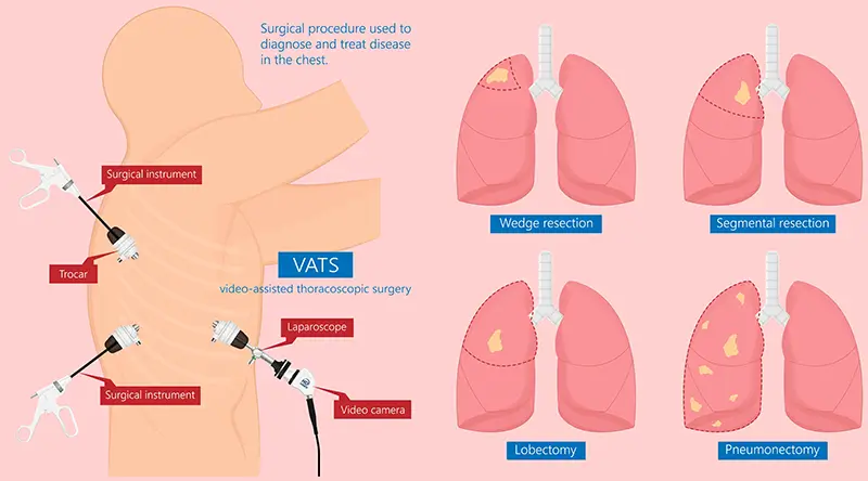
VATS can be used for procedures including:
- Lobectomy
- Anatomical segmentectomy
- Wedge resection
- Lung biopsy
- Pleurodesis
- Bullectomy
- Decortication
- Thymectomy
- Selected mediastinal procedures
Uniportal VATS
Uniportal VATS represents a further evolution of thoracoscopic surgery.
Instead of using multiple ports, the camera and surgical instruments are introduced through a single intercostal incision.
In experienced hands, uniportal VATS can be used for major anatomical pulmonary resections and other complex thoracic procedures.
The single-incision approach reduces the number of intercostal access points and may reduce chest-wall trauma and postoperative discomfort. However, the magnitude of these potential clinical benefits compared with multiportal VATS continues to be investigated and may vary according to the procedure and patient.
Robot-Assisted Thoracic Surgery (RATS)
Robotic-assisted thoracic surgery represents another major development in minimally invasive surgery.
The surgeon operates from a robotic console, controlling highly articulated instruments positioned inside the thoracic cavity.
Modern robotic platforms provide several technical capabilities:
- High-definition three-dimensional visualization
- Magnified surgical view
- Articulated instruments with a wide range of motion
- Tremor filtration
- Precise dissection in confined anatomical spaces
- Improved surgeon ergonomics
These characteristics can be particularly useful when performing delicate dissection around pulmonary vessels, bronchi, lymph nodes, and mediastinal structures.
RATS is currently used for procedures including:
- Lobectomy
- Anatomical segmentectomy
- Thymectomy
- Mediastinal tumor resection
- Selected complex pulmonary resections
- Selected esophageal and airway procedures
Uniportal Robotic-Assisted Thoracic Surgery (U-RATS)
One of the most recent and specialized developments in minimally invasive thoracic surgery is Uniportal Robotic-Assisted Thoracic Surgery (U-RATS).
Conventional robotic thoracic surgery was developed using multiple ports. U-RATS applies robotic technology through a single intercostal surgical access, combining the philosophy of uniportal surgery with the technological capabilities of robotic-assisted surgery.
The approach allows the surgeon to use three-dimensional high-definition visualization and articulated robotic instruments while limiting surgical access to a single incision.
U-RATS is currently performed by a limited number of highly specialized thoracic surgery teams internationally.
The technique requires advanced experience in minimally invasive thoracic surgery, robotic surgery, and uniportal techniques, and has a significant learning curve.
In experienced hands, U-RATS can be applied to increasingly complex procedures, including:
- Lobectomy
- Anatomical segmentectomy
- Thymectomy
- Mediastinal tumor resection
- Selected complex pulmonary resections
Potential advantages of the uniportal robotic approach include:
- A single surgical incision
- Fewer intercostal access points
- Three-dimensional magnified visualization
- Robotic instrument articulation and precision
- Potential reduction in chest-wall trauma and postoperative discomfort
- Excellent cosmetic results
- Early mobilization
- Potential for rapid postoperative recovery
Although the early published experience with U-RATS is encouraging, the available evidence remains less extensive than that for established VATS and multiportal RATS. Further comparative and long-term studies are required to determine whether reducing robotic surgery to a single access produces measurable clinical advantages beyond those already achieved with established minimally invasive approaches.
Open Thoracotomy vs. VATS vs. RATS
The different surgical approaches should not be considered competing techniques in every clinical situation. Each has advantages, limitations, and specific indications.
| Feature | Open Thoracotomy | VATS | RATS |
|---|---|---|---|
| Surgical access | Larger incision | One or more small incisions | One or more small incisions depending on technique |
| Rib spreading | May be required | Generally avoided | Generally avoided |
| Visualization | Direct surgical view | High-definition camera | Magnified 3D high-definition view |
| Instruments | Larger surgical access | Uniportal or multiportal | Uniportal or multiportal |
| Postoperative recovery | Generally longer | Generally faster | Generally faster |
| Hospital stay | Generally longer | Potentially shorter; selected patients may be discharged POD 1 | Potentially shorter; selected patients may be discharged POD 1 |
| Main role | Selected complex cases or when minimally invasive surgery is inappropriate | Broad range of thoracic procedures | Broad range of thoracic procedures, including complex minimally invasive surgery |
Importantly, outcomes depend not only on the surgical platform but also on patient selection, procedure complexity, perioperative protocols, and surgeon/team experience.
Conversion from a minimally invasive procedure to thoracotomy should not automatically be regarded as a surgical failure. When required because of unexpected bleeding, difficult anatomy, severe adhesions, or oncological considerations, conversion represents an appropriate decision made in the interest of patient safety.
Conditions Treated With Minimally Invasive Thoracic Surgery
Lung Cancer
Minimally invasive surgery has become an important component of treatment for resectable non-small-cell lung cancer.
Depending on tumor size, location, stage, pulmonary reserve, and other oncological characteristics, surgical options may include:
- Lobectomy
- Anatomical segmentectomy
- Wedge resection in appropriately selected situations
- Pneumonectomy in selected cases
The increasing role of anatomical segmentectomy
One of the most significant recent changes in lung cancer surgery has been the increasing role of anatomical segmentectomy.
Historically, lobectomy was considered the standard anatomical resection for most operable early-stage lung cancers.
Modern randomized trials, particularly JCOG0802/WJOG4607L and CALGB/Alliance 140503, have changed this paradigm.
These studies demonstrated that appropriately selected patients with small peripheral early-stage NSCLC can be treated with parenchymal-sparing resections without compromising key oncological outcomes compared with lobectomy.
The Japanese JCOG0802/WJOG4607L trial specifically demonstrated significant survival results for anatomical segmentectomy in patients with peripheral NSCLC ≤2 cm, although local recurrence was higher following segmentectomy.
Patient selection therefore remains essential.
The objective of modern precision lung surgery is not simply to remove less lung, but to achieve adequate oncological resection while preserving as much healthy pulmonary parenchyma as safely possible.
Pulmonary Nodules
The increasing use of CT and low-dose CT screening has led to the detection of increasingly small pulmonary nodules and ground-glass opacities.
When imaging and other diagnostic techniques cannot establish the nature of a suspicious lesion, surgical resection may be indicated for diagnosis and treatment.
Minimally invasive techniques can be combined with modern localization methods to accurately identify very small or deep pulmonary lesions.
Pleural Disease
Thoracoscopic surgery can help diagnose and treat:
- Pleural effusions
- Pleural tumors
- Empyema
- Pleural infection
- Recurrent pneumothorax
- Malignant pleural effusion
Procedures may include pleural biopsy, decortication, and different forms of pleurodesis.
Pneumothorax
Surgical treatment may be appropriate for certain patients with recurrent spontaneous pneumothorax, persistent air leak, or other established indications.
Depending on intraoperative findings and surgical strategy, treatment may include resection of identifiable blebs or bullae together with a procedure intended to reduce recurrence, such as mechanical or other forms of pleurodesis or pleural intervention.
The precise technique should be individualized.
Mediastinal Tumors
Minimally invasive surgery has become particularly important in mediastinal surgery.
Selected:
- Thymomas
- Thymic lesions
- Mediastinal cysts
- Neurogenic tumors
- Other mediastinal masses
Can be approached using VATS or robotic surgery.
Robotic articulation and three-dimensional visualization can be particularly useful in the confined anatomical space of the mediastinum.
Uniportal robotic surgery represents a further evolution of this approach in selected patients and experienced centers.
What to Expect Before, During and After Surgery
Before Surgery
Preoperative assessment is individualized according to the planned operation and patient.
It may include:
- Blood tests
- Electrocardiogram
- Pulmonary function testing
- CT
- PET-CT when indicated
- Bronchoscopy when indicated
- Cardiovascular assessment when appropriate
- Anesthesia assessment
Smoking cessation is strongly encouraged before surgery.
Prehabilitation, nutritional optimization and physical exercise may also form part of preparation for major thoracic surgery.
During Surgery
Most major minimally invasive thoracic procedures are performed under general anesthesia.
For many pulmonary resections, one-lung ventilation provides the surgical team with adequate intrathoracic working space.
The duration of surgery varies considerably according to the operation and its complexity.
A chest drain is commonly placed at the end of pulmonary surgery, although modern protocols increasingly aim to reduce the duration of chest drainage and, in selected cases, avoid postoperative drainage altogether.
Recovery and Hospital Stay
Modern thoracic surgery places increasing emphasis on Enhanced Recovery After Surgery (ERAS) principles.
These include:
- Multimodal analgesia
- Reduction of opioid requirements
- Regional analgesia when appropriate
- Early mobilization
- Early oral intake
- Optimized intravenous fluid management
- Early removal of urinary and intravenous catheters
- Optimized chest-drain management
Patients are encouraged to begin mobilizing as soon as clinically appropriate, frequently on the day of surgery or the following morning.
Hospital stay varies according to the procedure, patient characteristics, and postoperative course.
Following minimally invasive thoracic surgery, selected patients may be discharged as early as the first postoperative day, while others require several days of hospitalization.
Early mobilization, multimodal pain control, and optimized chest-drain management are integrated into perioperative care to facilitate safe early discharge whenever clinically appropriate.
After thoracotomy, hospital admission is generally longer, although modern ERAS pathways have also substantially improved recovery after open surgery.
Benefits of Minimally Invasive Thoracic Surgery
Compared with conventional thoracotomy, minimally invasive approaches have been associated with several potential benefits in appropriately selected patients, including:
- Smaller surgical incisions
- Avoidance of rib spreading
- Reduced postoperative pain
- Reduced blood loss in many procedures
- Earlier mobilization
- Shorter hospital stay
- Reduced respiratory morbidity in selected populations
- Faster functional recovery
- Improved cosmetic results
For appropriately selected patients undergoing lung cancer surgery, minimally invasive approaches can provide oncological outcomes comparable to open surgery while improving several aspects of postoperative recovery.
However, the objective of minimally invasive surgery should never be to achieve a smaller incision at the expense of surgical safety or oncological principles.
Risks and Limitations
Minimally invasive thoracic surgery remains major surgery.
Potential complications vary according to the procedure and may include:
- Bleeding
- Prolonged air leak
- Pneumonia
- Infection
- Atrial fibrillation or other cardiac rhythm disturbances
- Venous thromboembolism
- Respiratory complications
- Injury to surrounding structures
- Need for reintervention
- Conversion to thoracotomy
Conversion to open surgery may occasionally be necessary because of bleeding, severe adhesions, unexpected anatomy, inability to safely visualize critical structures, or oncological considerations.
Advances Shaping the Future of Thoracic Surgery
New Robotic Platforms
Robotic technology continues to evolve rapidly.
Newer systems aim to provide increasingly sophisticated visualization, greater instrument flexibility, smaller platforms, and potentially more efficient operating-room integration.
The development of uniportal robotic surgery reflects the broader trend toward combining increasingly sophisticated technology with progressively less invasive surgical access.
Artificial Intelligence and Surgical Planning
Artificial intelligence is increasingly being investigated across thoracic medicine and surgery.
Potential applications include:
- Pulmonary nodule detection
- Imaging analysis
- Risk prediction
- Three-dimensional anatomical reconstruction
- Surgical planning
- Intraoperative decision support
These technologies should be regarded as tools supporting clinical decision-making rather than replacing surgical judgment.
Advanced Imaging and Image-Guided Surgery
Image-guided techniques are increasingly important as lung cancer screening detects smaller and less palpable lesions.
Current technologies include:
- Indocyanine green (ICG) fluorescence
- Three-dimensional CT reconstruction
- Navigational bronchoscopy
- Robotic bronchoscopy
- Dye localization
- Microcoil localization
- Intraoperative cone-beam CT
- Hybrid operating-room techniques
ICG fluorescence can also assist during anatomical segmentectomy by helping identify the intersegmental plane.
These technologies are increasingly integrating diagnosis, localization, and surgical treatment.
Precision Lung Resection
Modern thoracic oncology is progressively moving away from a philosophy based simply on removing the largest possible amount of lung tissue.
The objective is increasingly precision oncological resection: removing the tumor with adequate margins and appropriate lymph-node assessment while preserving healthy pulmonary parenchyma whenever oncologically appropriate.
Anatomical segmentectomy, advanced imaging, and minimally invasive surgery are central to this development.
Non-Intubated and Tubeless Thoracic Surgery
Selected specialized centers are investigating strategies to further reduce perioperative invasiveness.
Non-intubated thoracic surgery allows selected procedures to be performed under spontaneous ventilation using carefully designed anesthetic and regional analgesic techniques.
Similarly, tubeless protocols aim to avoid or shorten the use of postoperative chest tubes, urinary catheters, and intravenous lines in carefully selected patients.
These approaches require careful patient selection and experienced multidisciplinary teams.
Frequently Asked Questions
Is robotic thoracic surgery better than VATS?
There is no single approach that is optimal for every patient.
Both VATS and RATS are established minimally invasive approaches that can achieve favorable outcomes.
Robotic surgery provides three-dimensional visualization and articulated instruments, while VATS is highly effective and widely established.
The optimal technique depends on the disease, procedure, patient anatomy, and, importantly, the expertise of the surgical team.
What is Uniportal RATS?
Uniportal RATS, or U-RATS, is robotic thoracic surgery performed through a single intercostal surgical access rather than the multiple ports traditionally used for robotic surgery.
It combines the principles of uniportal thoracic surgery with the visualization and articulated instrumentation of robotic technology.
It remains a specialized technique performed by a limited number of experienced thoracic surgical teams internationally.
Is U-RATS better than conventional robotic surgery?
Current findings support U-RATS as a safe and feasible approach in experienced hands, but comparative evidence remains limited.
The reduction to a single surgical access is conceptually attractive and may reduce intercostal trauma, but further studies are needed to establish whether it provides clinically significant advantages over multiportal RATS or established VATS techniques.
Can patients really go home the day after thoracic surgery?
Yes, selected patients.
Modern minimally invasive techniques combined with ERAS protocols, effective pain management, early mobilization, and optimized chest-drain management can allow discharge as early as the first postoperative day.
However, discharge should always depend on clinical recovery and established safety criteria rather than on achieving a predetermined length of stay.
Is minimally invasive surgery suitable for lung cancer?
Yes. Minimally invasive surgery is widely used for appropriately selected patients with resectable lung cancer.
Depending on tumor characteristics and patient factors, procedures may include lobectomy, anatomical segmentectomy, or other appropriately selected resections.
The surgical approach must always respect the same oncological principles regardless of incision size.

Dr. Régulo José Ávila Martínez is a specialist in Thoracic Surgery at Hospital Ruber Internacional and Hospital Universitario La Luz in Madrid.
After graduating in Medicine and Surgery in 2006, he undertook specialist training in Thoracic Surgery through the Spanish MIR system. Dr. Martínez later earned his Doctorate from the Complutense University of Madrid in 2017 in Medicine and Surgery. His professional development has included advanced training in thoracic oncology and clinical experience in Spain and Germany.
His clinical practice focuses particularly on minimally invasive thoracic surgery, including video-assisted and robotic-assisted approaches and advanced uniportal techniques.
He is also involved in teaching and research in thoracic medicine and surgery.
His distinctions include the Cross of Merit with White Distinction, the Balmis Operation Medal, and an Award for Best Communication in Thoracic Surgery.

Hospital Ruber Internacional, part of the Quirónsalud hospital group in Madrid, provides multidisciplinary care across multiple medical and surgical specialties.
Its surgical technology includes the Da Vinci Xi robotic surgical system, enabling robotic-assisted procedures across several specialties.
Within thoracic surgery, advanced minimally invasive approaches and the integration of robotic technology, advanced imaging, modern anesthesia, and enhanced-recovery pathways support an increasingly personalized and minimally invasive approach to thoracic surgical care. At the Madrid center, the minimally invasive thoracic surgery program includes Uniportal Robotic-Assisted Thoracic Surgery (U-RATS), which is used for selected anatomical pulmonary resections and mediastinal procedures.
References:
Featured Blogs

Medically Induced Coma: What It Is, How It Works & Recovery