
Bullous Pemphigoid: Causes & Risk Factors, Symptoms, Diagnosis, and Treatment

Medically Reviewed by Dr. Sony Sherpa, (MBBS) - July 28, 2025
Bullous pemphigoid is a skin disorder caused by an autoimmune response, resulting in the development of large, tense blisters. Predominantly seen in older adults, it can impair quality of life through persistent itching and skin irritation.
This blog aims to provide a comprehensive overview of bullous pemphigoid, explaining its causes, symptoms, diagnosis, treatment options, and prognosis, with the goal of raising awareness and supporting individuals affected by this condition in gaining a better understanding of it.
What is Bullous Pemphigoid?
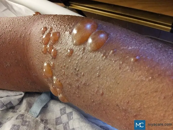
Bullous pemphigoid is the most prevalent autoimmune blistering disorder affecting the subepidermal layer of the skin. It primarily affects older adults, particularly those aged 60 to 80, with a slight predominance in females. It is characterized by the development of large, fluid-filled blisters (bullae), typically on flexural areas such as the inner thighs, underarms, and lower abdomen. However, extensor surfaces such as the arms and legs can also be involved. Lesions are symmetrical in distribution in many cases. These blisters are usually tense and do not rupture easily, and are often accompanied by redness, itching, and inflammation. The estimated annual incidence ranges from 2 to 22 cases per million in the general population, depending on geographic and demographic factors. Although considered rare, its impact can be significant in elderly individuals due to a heightened risk of complications from blisters, infections, and treatment side effects. Recent population-based studies indicate that the incidence of bullous pemphigoid is rising, likely due to increased awareness and an aging global population.
Types of Bullous Pemphigoid
There are several clinical variants of bullous pemphigoid:
- Classic Bullous Pemphigoid: Presents with widespread, tense blisters on normal or reddened skin.
- Urticarial Pemphigoid: Appears as intensely itchy, hive-like rashes before blisters develop.
- Localized Bullous Pemphigoid: Limited to specific body regions, such as the lower legs.
- Pemphigoid Gestationis: A rare pregnancy-related form, usually occurring in the second or third trimester.
Causes and Risk Factors
Bullous pemphigoid is primarily an autoimmune disease with an idiopathic origin in most cases, meaning the exact cause remains unknown. However, several environmental, immunological, and genetic factors have been associated with its onset, particularly in elderly individuals.
Idiopathic Nature and Immune Dysregulation
In bullous pemphigoid, the body’s immune system mistakenly attacks components of the skin's basement membrane, specifically the BP180 (type XVII collagen) and BP230 proteins that help anchor the outer layer of skin (epidermis) to the underlying dermis. This autoimmune response leads to the accumulation of inflammatory cells and the formation of subepidermal blisters, which separate the top layer of skin from the underlying tissue.
The immune attack involves IgG autoantibodies and complement activation, resulting in the disruption of the hemidesmosomes, which are key anchoring structures in the skin. This separation causes the skin to lift, forming the classic blisters seen in the disease. The initial immune dysregulation is often spontaneous, especially in older adults.
Age
One of the most significant risk factors is advanced age. Most cases occur in individuals over 60 to 80 years of age, with incidence increasing with age. Age-related immune senescence may contribute to the breakdown of self-tolerance and trigger autoimmune responses.
Neurological Disorders
There is a well-established association between bullous pemphigoid and certain neurological diseases, including:
- Parkinson’s disease
- Alzheimer’s disease
- Multiple sclerosis
- Stroke
This link may involve shared antigens between the nervous system and skin or generalized immune dysregulation.
Drug-Induced Bullous Pemphigoid
Certain medications have been implicated in triggering or unmasking bullous pemphigoid. This includes:
- Diuretics
- Antibiotics
- Antipsychotics
- Dipeptidyl peptidase-4 inhibitors (DPP-4 inhibitors) used in type 2 diabetes
These cases are termed drug-induced bullous pemphigoid and may resolve after discontinuation of the offending agent, though immunosuppressive therapy is still often required.
Genetic Predisposition
Although not inherited, bullous pemphigoid may occur more frequently in individuals with certain HLA genotypes, suggesting that genetic susceptibility may be a factor in disease development.
Other Skin Conditions and Triggers
Physical trauma, surgical wounds, burns, UV exposure, and radiotherapy can act as triggers in genetically predisposed individuals. Pre-existing inflammatory skin diseases, like eczema or psoriasis, may also increase vulnerability.
Pregnancy-Associated Bullous Pemphigoid: Pemphigoid Gestations
Pemphigoid gestationis is a rare variant that occurs in pregnant women, usually during the second or third trimester. It is caused by similar autoantibodies and may flare postpartum or with future pregnancies. Though uncommon, it highlights the role of hormonal and immune changes in disease expression.
Bullous Pemphigoid Symptoms
Bullous pemphigoid symptoms can vary widely between individuals, but they typically follow a progression from itchy, nonspecific skin changes to the development of tense, fluid-filled blisters. The symptoms can be distressing and may significantly impact quality of life, particularly in the elderly.
Early Symptoms
The disease often begins with:
- Itching (pruritus), which may be intense
- Urticarial (hive-like) or eczematous lesions
- Red, inflamed patches of skin without visible blisters
This stage is frequently misdiagnosed as dermatitis or allergic reactions, delaying accurate diagnosis.
Blister Formation
As the disease progresses, classic bullous lesions appear:
- Tense bullae (blisters) that are filled with clear or blood-tinged fluid
- Often arise on non-inflamed or mildly reddened skin
- May be localized (e.g., lower legs) or widespread
Blisters typically do not rupture easily due to their subepidermal origin, distinguishing them from more fragile blisters seen in conditions like pemphigus vulgaris.
After Blisters Break
When bullae rupture, they leave behind:
- Erosions or open sores
- Crusting or scabbing as the skin begins to heal
Skin tearing can occur, especially in frail or elderly skin, increasing the risk of secondary infection.
Mucous Membrane Involvement
Though less common compared to pemphigus vulgaris, some patients may have mucosal involvement, including:
- Mouth (bullous pemphigoid in the oral cavity) – causing pain, burning, or difficulty eating
- Nasal mucosa
- Throat – may lead to hoarseness or difficulty swallowing
- Genitals – resulting in painful ulcers or erosions
Mucosal lesions are usually shallow and erosive, and may be overlooked unless specifically examined.
Other Symptoms
- Fatigue due to chronic inflammation or disrupted sleep from itching
- Pain if lesions become infected
- Dehydration in severe and widespread blistering
Complications
- Secondary bacterial infections (e.g., impetigo, cellulitis)
- Sepsis, especially in immunocompromised or elderly individuals
- Electrolyte imbalance and fluid loss in extensive disease
- Scarring and pigmentation changes after healing
- Functional decline, particularly in older adults, due to limited mobility or pain
Patients with extensive disease may also face psychological distress or depression due to the chronic and visible nature of the skin condition.
Diagnosis
Bullous pemphigoid is diagnosed through a combination of clinical assessment and specialized laboratory tests.
- Physical examination and symptom review: Doctors assess skin lesions, noting the presence of tense blisters typically on the flexural areas, and check for mucous membrane involvement. Itching and rash history are important clues.
- Skin biopsy and direct immunofluorescence test: A small sample of affected skin is taken to be examined under a microscope. The biopsy shows subepidermal blisters with inflammatory cells. Direct immunofluorescence reveals linear deposits of IgG and C3 along the basement membrane zone, confirming the autoimmune attack on skin structures.
- Blood tests: Serum is tested for circulating bullous pemphigoid antibodies, mainly targeting BP180 and BP230 proteins.
What is the Nikolsky Sign in Bullous Pemphigoid?
The Nikolsky sign is a clinical test where gentle lateral pressure on the skin causes the epidermis to shear off, indicating skin fragility.
- In bullous pemphigoid, the Nikolsky sign is typically negative because the blisters are subepidermal and tense, meaning the skin is more resistant to shearing.
- This contrasts with pemphigus vulgaris, where the Nikolsky sign is usually positive due to fragile, superficial blisters.
Differential Diagnosis of Bullous Pemphigoid
Several skin conditions mimic bullous pemphigoid, so accurate diagnosis is essential.
Bullous Pemphigoid vs Pemphigus Vulgaris
- Bullous pemphigoid features tense, subepidermal blisters, mainly in elderly patients, with rare mucosal involvement and a negative Nikolsky sign.
- Pemphigus vulgaris causes flaccid, easily ruptured blisters often involving mucous membranes and a positive Nikolsky sign. Histology and immunofluorescence differentiate them.
Bullous Impetigo vs Bullous Pemphigoid
- Bullous impetigo is a superficial bacterial infection, mostly in children, with fragile blisters and pustules, and responds well to antibiotics.
- Bullous pemphigoid is autoimmune and requires immunosuppressive treatment.
Other Conditions to Differentiate
- Epidermolysis bullosa: Inherited blistering starting from infancy.
- Shingles (Herpes Zoster): Painful, dermatomal rash with vesicles.
- Cicatricial pemphigoid: Chronic mucous membrane scarring.
- Stevens-Johnson Syndrome (SJS): Acute mucocutaneous reaction with systemic symptoms.
Can Bullous Pemphigoid Occur in Patients with Lymphedema?
Bullous pemphigoid is primarily characterized by tense blisters rather than diffuse swelling. While uniform swelling alone is not typical of BP, cases have been reported in which localized BP develops in areas affected by lymphedema.
Bullous Pemphigoid Treatment
Medications
Topical corticosteroids are effective for mild or localized disease. Oral corticosteroids are used for moderate to severe cases to reduce inflammation. Immunosuppressive drugs help minimize steroid use and control chronic disease.
Biologic therapies, notably dupilumab (Dupixent), have shown promising results in refractory cases by targeting immune pathways. This was approved by the FDA for the treatment of Bullous Pemphigoid in June 2025.
Supportive Care
Wound care involves gentle cleansing and infection prevention with antiseptics and non-adhesive dressings. Antihistamines help relieve intense itching. Emollients restore skin moisture and barrier function, reducing irritation.
Lifestyle Tips and Management
Home remedies include avoiding known triggers such as trauma or irritants and practicing gentle skin care with mild soaps and lukewarm water. Fall prevention is crucial in elderly patients due to fragile skin and blister pain. Additionally, ensuring safe environments and mobility aids reduces injury risk.
Nutritional support emphasizes avoiding processed foods, excessive sugars, and irritants while promoting an anti-inflammatory diet rich in omega-3 fatty acids, antioxidants, and vitamin D. Hydrating foods and probiotics may support skin and immune health. Patients can shower safely with lukewarm water and gentle cleansers, followed by immediate moisturizing, avoiding hot water and vigorous scrubbing.
Prognosis and Long-Term Outlook
Bullous pemphigoid is a chronic but manageable condition. Healing time varies. With treatment, active blistering often improves within weeks to months, but complete remission may take longer. Flare-ups can occur, and some patients achieve long-term remission. Long-term medication use, especially corticosteroids and immunosuppressants, may cause side effects such as osteoporosis, infections, and diabetes, making regular follow-ups and medication monitoring essential.
Although rarely fatal, bullous pemphigoid can be life-threatening in elderly or immunocompromised patients due to complications, such as infections. The mortality rate is higher compared to the general population, but timely treatment can improve outcomes.
When to See a Doctor
Persistent itching or unexplained blisters, especially when accompanied by sudden changes in the skin, should prompt medical evaluation. Older adults should be particularly vigilant, as early diagnosis improves management and reduces the risk of complications.
Frequently Asked Questions
Do COVID-19 vaccines cause bullous pemphigoid?
There are rare reports of bullous pemphigoid onset or flare-ups after COVID-19 vaccination, but the benefits of vaccines outweigh the risks. Consult your doctor.
Can diabetes cause bullous pemphigoid?
Diabetes itself does not cause bullous pemphigoid, but certain diabetes medications (like DPP-4 inhibitors) may trigger it in susceptible individuals.
What is a bullous disease of the esophagus?
Bullous diseases can rarely affect the esophagus, causing blisters and erosions, leading to swallowing difficulties.
Can bullous pemphigoid affect the eyes?
Though uncommon, mucous membrane involvement can include the conjunctiva, causing redness and irritation.
Is bullous pemphigoid related to cancer?
Bullous pemphigoid is generally not linked to cancer but requires evaluation to exclude paraneoplastic causes if suspected.
Is bullous pemphigoid associated with dementia?
There is an observed association between bullous pemphigoid and neurological diseases, including dementia, possibly due to shared immune mechanisms.
To search for the best Dermatology Healthcare Providers in Croatia, Germany, Greece, India, Malaysia, Singapore, Slovakia, Spain, Thailand, Turkey, Ukraine, the UAE, UK, the USA, please use the Mya Care search engine.
The Mya Care Editorial Team comprises medical doctors and qualified professionals with a background in healthcare, dedicated to delivering trustworthy, evidence-based health content.
Our team draws on authoritative sources, including systematic reviews published in top-tier medical journals, the latest academic and professional books by renowned experts, and official guidelines from authoritative global health organizations. This rigorous process ensures every article reflects current medical standards and is regularly updated to include the latest healthcare insights.

Dr. Sony Sherpa completed her MBBS at Guangzhou Medical University, China. She is a resident doctor, researcher, and medical writer who believes in the importance of accessible, quality healthcare for everyone. Her work in the healthcare field is focused on improving the well-being of individuals and communities, ensuring they receive the necessary care and support for a healthy and fulfilling life.
References:
Featured Blogs

Medically Induced Coma: What It Is, How It Works, Who Benefits, and Recovery Outcomes

