Digestive System Overview

Medically Reviewed by Dr. Sony Sherpa (MBBS) - September 12, 2024
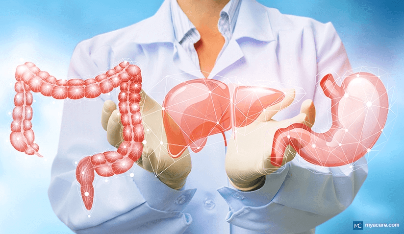
The digestive system is one of the largest organ systems in the body, as well as one of the most crucial for its overall functioning.
The digestive tract refers largely to the alimentary canal, a hollow organic tube that connects the mouth to the anus and that facilitates digestion with the aid of the digestive organs that branch off of it.
The below overview describes the functions of the digestive system, the organs involved in digestion and absorption as well as how these organs are regulated.
Functions of the Digestive System
Each time any event occurs within the body, it occurs primarily as a series of chain chemical reactions at the micro (cellular) level, which in turn propels chain events at the macro (organ tissue) level. The digestive system is the chief system in place to ensure that the body receives an adequate supply of basic building blocks in order to sustain these chemical reactions. Subsequently, these reactions produce further substances that are absolutely required for optimal biological function and maintenance, such as energy production and cellular repair.
In order to achieve continued homeostasis, the functions of the digestive system can be divided into four main categories that are equally vital to the survival of any complex organism:
- Digestion. Digestion is the process of breaking food down into smaller and smaller units until the units are able to be absorbed into the body. Most of the cells and bacteria in the digestive tract are involved in breaking food down. Food is typically transformed from its whole state into four main macronutrient categories: carbohydrates, proteins, sugars, and fats. Fiber aids digestion by facilitating bacterial secretory activity and stool formation. Macronutrients are either accompanied by or broken down into micronutrients in the diet, which consist of minerals, vitamins, and other antioxidant compounds such as plant polyphenols (flavonoids, tannins, etc).
- Absorption. After food has been broken down and prepared into smaller molecules, the cells of the gut can then take them up, and after further processing, transport them to where they are required via the bloodstream. This is the process of absorption. All nutrients have differing ways of being absorbed and are generally taken up at unique portions of the digestive tract in line with the respective processes of digestion relevant to the area. For example, the liver and gallbladder secrete enzymes that break down fats and proteins, and those enzymes are also typically required for their uptake into cells.
- Excretion. The digestive system is involved in both waste elimination and excretion. Waste from circulation passes through the liver and is emptied into the gastrointestinal tract for removal from the body. These waste products are thus eliminated, made to join incompletely digested food particles moving along the tract for further bacterial refinement and eventual excretion.
- Protection. Like the skin, the organs in the digestive tract are exposed to the outer environment and form part of our frontline defense system against foreign threats. The gut’s intricate system of defense works in tandem with the other described functions, ensuring that hazardous material is broken down alongside food, that only the correct nutrients are absorbed, and that all dangerous media is excreted alongside waste. As a result, immune function and gut function are closely linked, and if one suffers, the other is bound to follow suit.
Digestive System Regulation
The process of digestion is facilitated by the movement of food through the gastrointestinal tract (GIT) as well as the release of important secretions along the way.
During digestion, food is propelled by two layers (three in the stomach) of smooth muscle tissue that line the entirety of the tract. The muscles contract to gently propel food until it is excreted, while the cells and bacteria lining the internal walls of the tract break food down and absorb nutrients. Certain accessory organs, such as the gallbladder and pancreas, secrete enzymes that promote digestion and absorption. [1]
The gastrointestinal movement of food is referred to as peristalsis[2].
Peristalsis and the enzymatic secretions of the gut are regulated by the nervous system, the endocrine system, the vascular system, and internally amongst intestinal cells themselves.
Intrinsic Gut Regulation of Peristalsis
The small intestine and large intestine rely on intrinsic regulation, carrying unique bundles of nerve fibers that inform the smooth muscle tissue therein to contract during peristalsis. If the gut were disconnected from the nervous system, it would still be able to perform peristalsis using its intrinsic regulatory mechanisms, yet the stomach and other digestive organs would fail to function without input from the nervous system.[3]
The intrinsic regulation of the gut works through localized conductance of electrical impulses through the gut’s nerve clusters. In response to these electrical impulses, the smooth muscle cells experience shifts in their membrane potentials, moving from a low to a high electrical potential. A high electrical potential causes an influx of calcium (Ca2+) into the cell, leading to a contraction - presuming there is a sufficient supply of Ca2+ and no interfering cellular activity that may hamper the process (e.g., localized gut inflammation).
The following segment of the tract tends to have a lower electrical potential than the portion that is busy contracting, allowing for it to contract immediately afterward as the electrical potential gradient moves from an area of high potential to one of low. In this way, continuous waves of contractions are coordinated along the entire tract while food is moving through, with each cell acting as a pacemaker that contributes toward the phenomenon of peristalsis.
Each contraction propels food approximately 5-20mm along the small intestine, which can be anywhere between 4 and 6m in length. Food typically takes 4 to 6 hours to pass through the stomach and small intestine (the longest part of the tract) and can take 36 hours[4] or longer to finish digesting in the large intestine before being excreted. The extra time is usually taken up by the final stages of digestion (fermentation) in the colon, yet any part of the process can be slowed down or sped up depending on the health of the individual.
The healthy gut takes between 2 and 5 days to digest a meal on average[5].
Neuro-Endocrine Regulation
The gut houses a sophisticated neuro-endocrine system of regulation in order to maintain proper function. This is especially true of the stomach and the accessory digestive organs such as the liver, gallbladder, and pancreas. The system is equally as complex as that seen in the brain, leading some scientists to classify the gut as a second brain or “little brain.”[6]
The central and peripheral nervous systems regulate digestion through the translation of nerve impulses into active hormones. These hormones have a wide range of actions, from maintaining satiety to stimulating digestive secretions.
Lastly, the organs themselves have additional endocrine cells that instruct the organ in response to localized signals at the cellular level. [7]
Peripheral Nervous System
The vagus nerve (referring to a bundle of nerve fibers) runs along the torso, including the entire digestive tract, and is largely involved in nervous regulation of the heart, lungs, and gut.
The parasympathetic nervous system governs the vagus nerve, and it is one of the main factors involved in the ‘rest and digest’ response. The stomach and many of the accessory organs rely on neuro-endocrine feedback from the vagus nerve in order to carry out processes during digestion and absorption.
When the sympathetic nervous system initiates a stress response (fight or flight response), the vagus nerve informs the gut to bring digestion to a halt. This is accompanied by less blood flow to the digestive tract and more blood flow to the periphery, less glucose uptake, and bodily conservation of cellular nutrients.
The peripheral nervous system also picks up motor and sensory information from all digestive organs, relaying information to the central nervous system and brain if any part of the tract is bloated or empty and promoting an appropriate response.
Central Nervous System
The central nervous system is connected to the peripheral nervous system via the spinal cord and influences digestion in a number of ways. Digestion is largely about the involuntary relaxation and contraction of abdominal muscles at the right place and at the right time. The nervous system helps to coordinate the relaxation and contraction of certain areas of the tract, such as the valve that releases partially digested food from the stomach into the intestines.
Aside from vagus nerve stimulation and motor information from specific digestive organs, the hypothalamus in the brain monitors the blood for any fluctuations associated with digestion (or starvation), such as hunger hormones, stress hormones, nutrient levels, and more. This part of the brain is also partly responsible for coordinating states of stress or relaxation, the latter of which is conducive to digestion and the former of which deactivates digestive processes.
The hindbrain (medulla oblongata) is also involved in regulating digestive function as far as involuntary reflexes are concerned. When the gut is mechanically distended (usually after eating a full meal), motor information travels to the medulla which responds by lowering heart rate (or contractility) and redirecting blood flow away from the affected area. Several portions of the hindbrain, such as the pons, are actively involved in regulating digestive function alongside the hypothalamus.
Multiple other areas of the brain are also involved in coordinating digestive functions, including the cerebellum, thalamus, amygdala, and several specialized nerve clusters in the cerebral cortex.[8]
Endocrine Cell Regulation
Digestive organs such as the pancreas, liver, and stomach all have their own unique system of hormonal communication at the cellular level. Neuroendocrine cells reside in all digestive organs and translate nerve signals from the nervous system into hormonal instructions that the cells in the organ can receive. Additionally to neuroendocrine cells, other endocrine cells exist that generate localized hormonal signals in response to chemical stimuli, ensuring that the organ functions optimally.
Autoregulation
Autoregulation refers to the regulation of blood flow in the arteries that are connected to an organ. As with all other organ tissues, autoregulation has a role to play in regulating activity in the gastrointestinal tract. This cardiovascular mechanism ensures that blood flow to the gut is adequate in supply, properly oxygenated, and that nutrients can be sufficiently absorbed.[9],[10]
How Digestion Works: A Brief Walk Through Gastrointestinal Anatomy
Digestion is similar all the way along the alimentary canal: food is propelled through, substances are secreted to aid digestion, and nutrients are harvested from transiting food molecules at nearly every step of the way. The process can be summed up in the following four stages[11]:
- The mouth and stomach refine the size of food particles into macromolecules through chemical and mechanical breakdown.
- The small intestine breaks macromolecules down into micronutrients and facilitates their absorption.
- The colon, with its rich probiotic density, ferments the remaining food particles, produces additional bacterially derived nutrients, and absorbs these nutrients as well as water.
- The rectum stores waste until it is ready for excretion.
The specific function of each part of the digestive tract is discussed in more depth below.
Mouth
Digestion begins in the mouth with food consumption. Salivary glands rest inside the oral cavity and produce salivary amylase (i.e., saliva), an enzyme that serves to break down carbohydrates and other larger molecules into smaller units.
The teeth are technically digestive organs that process food into an acceptable format for the stomach. Once food has been chewed and swallowed, it starts to form a bolus that evolves through the course of digestion.
Stomach
The mouth is connected via the esophagus to the top part of the stomach. The walls of the stomach consist of three thick layers of smooth muscle that are suggestive of the organ’s role in digestion.
When the bolus enters the stomach, the top portion of the organ enlarges, which is also partly why the belly distends when one eats. An empty stomach is the size of one’s fist yet it has the ability to expand up to 75 times that size to accommodate excessive food consumption. The muscular sides of the stomach mechanically knead the bolus, refining the work of the teeth.
Simultaneously, cells in the stomach walls produce acids that chemically break the food down. These acids are strong enough to digest the walls of the stomach. Thus, the stomach is protected by a layer of bicarbonate-rich (acid-resistant) mucous referred to as the mucosa. The lining of the stomach is shed and turns over every 3 to 6 days.
In the gastric mucosa, deep pores can be seen that descend into gastric glands, lined with cells that secrete gastric juices. The majority of gastric glands produce mucous, with different sections of the stomach wall producing appropriate digestive juices. The gastric glands are home to neuro-endocrine cells that signal for other cells inside the gland to produce stomach acids and enzymes, including[12]:
- Gastrin is a hormone that is released by gastric endocrine cells the moment the stomach senses the breakdown of proteins, which are present in most foods and some drinks. Gastrin-producing cells mostly reside at the bottom of the stomach. When gastrin is present, it signals for the stomach to empty into the intestine, relaxing the valve and promoting intestinal peristalsis.
- Histamine is produced by the stomach to stimulate the production of hydrochloric acid.
- Hydrochloric acid (HCL) is a very strong acid that is able to reduce chewed food into a soft paste known as chyme. It is secreted by parietal cells inside gastric glands. Aside from being a strong digestive enzyme in its own right, HCL is required to activate pepsin.
- Pepsin (from pepsinogen) is a potent digestive enzyme that works in tandem with HCL to break food down into chyme. Chief cells in gastric glands secrete pepsinogen, which is transformed into pepsin in the presence of HCL.
- Lingual Lipase breaks down fats in the stomach, specifically triglycerides into free fatty acids and simpler fats (mono- and diglycerides).
- Intrinsic Factor is another enzyme secreted by gastric parietal cells that is essential to digestion in humans. This enzyme allows for the absorption of vitamin B12 in the small intestine, which is entirely vital for numerous bodily functions including blood cell formation and nerve cell activity.
These chemical agents, in combination with the mechanical churning of the stomach walls, dissolve the bolus into a highly acidic paste known as chyme. Chyme comprises macromolecules as opposed to the larger particles that are still present in chewed food.
Once the food has been turned into chyme, it can be released into the small intestine, where nutrients are extracted and absorbed. Certain substances pass through to the bloodstream from the stomach, being ferried off via the spleen into the liver for instant processing. These include caffeine and alcohol.
The gastric phase of digestion can last anywhere between 3 and 4 hours. Most of this phase is taken up by gastric emptying. Not all the contents can be released at once into the gut for processing, or this would create major problems with digestive acidity. In this respect, the stomach also acts as a storage organ that sets the pace for peristalsis in the gut, only releasing small amounts of chyme at a time into the small intestine. As the small intestine takes a longer time to process high-fat foods, fatty chyme can remain in the stomach for up to 6 hours or longer.
Through the nervous system (specifically the vagus nerve), signals from the intestines, brain, and stomach coordinate the release or inhibition of gastric juices. When under emotional stress or when the intestine is already full of chyme, the stomach receives signals that ensure it stops producing digestive enzymes.
In the reverse scenario, when the gut is empty or when we see food that we want to eat, the brain signals to the stomach to release gastric juices as well as the hunger hormone, ghrelin. When we are full, another hormone, leptin, serves to counter the effects of ghrelin and induce a feeling of satiety.
Spleen
The spleen is technically a part of the lymphatics system, the organ system associated with the immune system. However, due to its very close position and connection to the stomach, the pancreas, and the liver, the spleen is being investigated for a potential role in the digestive system - which is likely given the evidence.[13]
The primary functions of the spleen relate to filtering blood, removing and disposing of any hazardous material, and acting as a storage reservoir for platelets and red blood cells. The spleen is also responsible for removing any faulty red blood cells from circulation, breaking them down, and recycling their basic components (such as iron) in order to facilitate further healthy blood cell production. [14]
In a healthy person, oxygenated blood moves from the spleen into the stomach via small arteries, while deoxygenated blood moves from the stomach into the spleen. In those with gastric disorders, this natural flow is reversed and appears to go hand-in-hand with pressure on the liver. When the liver is over-burdened or under-functioning, deoxygenated blood can flow back into the spleen and, subsequently, the stomach, creating an excess of waste products that would have normally been sent to the liver for appropriate processing.
In this context, the spleen likely serves as a secondary filtration system for the stomach before the blood flow reaches the liver, and may additionally have endocrine and immunological actions on the stomach via unknown secretory actions. Historically, traditional doctors and complementary practitioners kept the spleen in mind when treating gastric conditions, noting that it improved the outcomes. More research is required to identify the potential role of the spleen in the digestive system.
Small Intestine
The small intestine is where the majority of nutrient absorption takes place. While the small intestine may have a smaller diameter than the large intestine, it is actually the longest portion of the digestive tract and can range anywhere between 4 and 6 meters in length on average.
The cells lining the small intestines have villi and microvilli, which are very small finger-like projections that continuously undulate (or ripple), aiding the movement of food through the gut over and above digestive contractions. Each of these villi absorbs nutrients and is supplied with capillaries that serve to transport the nutrients toward the liver for further processing and eventual bodily dissemination. Villi are attached to cells lining the tract, which also secrete enzymes to help with both digestion and absorption.
Collectively, the villi form the brush border of the intestinal tract and their enzymes are known as brush border enzymes. Brush border cells can have as many as 3000 microvilli covering their surfaces, promoting efficient nutrient absorption. The small intestine absorbs the largest variety of nutrients from food, including proteins, fats, carbohydrates (sugars), vitamins, minerals (including electrolytes), water and other micronutrients.[15]
Secretory cells in the small bowel help to keep the mucosa of the intestines intact due to their alkaline secretions.
The small intestine is anatomically divided into the following three segments:
- Duodenum
- Jejunum
- Ileum
The duodenum is the first portion of the small intestine and also the shortest segment, averaging 20-25cm or 7.9-9.8 inches.
While it is the shortest portion of the small intestine, the duodenum is vital for completing digestion in the small intestine and promoting optimal nutrient uptake. It receives chyme from the stomach, as well as secretions from the pancreas and bile acids from the liver and gallbladder, all of which further aid digestion.
When the duodenum receives chyme, it secretes its own gastrin, which stimulates the production of more gastric juices from the stomach that promote its emptying. As soon as the duodenum becomes distended with chyme, stretch receptors signal for gastrin production to stop and for the stomach’s lower valve to close.
Throughout the duodenum reside Brunner glands, which produce bicarbonate in order to raise the pH and neutralize the excessive acidity of gastric secretions. The alkaline environment of the duodenum deactivates pepsin and other gastric juices, enabling alternative enzymes to come alive in the higher pH, such as those secreted from the pancreas.
The other sections of the tract, the jejunum (2.5m or 8.2ft) and the ileum (3m or 9.8ft), are responsible for absorbing nutrients from the chyme after it has been altered by the duodenum, housing trillions of microvilli. Fats, sugars, and proteins are absorbed in the jejunum. Any leftover nutrients, vitamin B12, and water are absorbed in the ileum. Bile is absorbed in this portion of the tract as well and is eventually recycled for use in the liver.[16]
Digestion of Proteins
Cholecystokinin (CCK) is a hormone released from the cells of the duodenum in response to proteins. This hormone stimulates for the pancreas to release enzymes that break proteins down (proteases). Proteases break proteins down into amino acids and oligopeptides.
An activating enzyme, enteropeptidase (or enterokinase), is then released from duodenal cells in order to activate the pancreatic proteases, which are initially secreted in their inactive forms. Once altered by proteases, protein peptides are the right shape and size for absorption along the tract.
Digestion of Fats
CCK also slows gastric emptying, which is a requirement for the digestion of fats. Lipases are enzymes that break fats down into smaller molecules, namely fatty acids, monoacylglycerol, and monoglycerides.
These enzymes originate from the mouth, stomach, and pancreas and need a longer time to work their actions. As a result, CCK is expressed more when the tract is processing fatty food, slowing digestion down and allowing for better fat breakdown.
Colipase, lipase, cholesterol ester hydrolase, and phospholipase A2 are secreted by the pancreas to initiate the breakdown of fats. Bile from the liver and gallbladder is secreted into the duodenum after the pancreas and is required for fat and protein digestion and absorption. Bile helps to emulsify fats and water in the gut, a requirement for the action of the water-soluble enzymes that break fat down.
Digestion of Carbohydrates
As with lipids and proteins, carbohydrates are broken down in the duodenum thanks to further secretions from the pancreas. Digestion of carbs only occurs in the mouth and duodenum, with chewing and salivary amylase being a vital first component of the process.
Carbohydrates are broken down from complex sugars (disaccharides) into simple sugars (monosaccharides). Complex sugars include maltose, lactose, and sucrose and are broken down to form glucose, galactose, and fructose, respectively.[17]
Digestion of Micronutrients
Macromolecules release micronutrients when they are broken down into basic building blocks. The acids from the stomach and enzymes in the duodenum aid in the process of micronutrient digestion, as do enzymes from probiotic bacteria in the gut.
Sodium, chloride, magnesium, potassium, calcium, phosphate, iron, and water are passively absorbed in the jejunum, ileum, and colon. These are commonly absorbed by similar chemical mechanisms involving hydrogen ions and acid-base chemistry. More micronutrients taken up by the small intestine include oxalates, water-soluble vitamins (vitamin C and B vitamins), fat-soluble vitamins (A, E, D, K), and plant-based compounds such as tannins and bioflavonoids.[18]
The Biliary System
The biliary system refers to the pancreas, liver, and gallbladder, the accessory digestive organs that empty their secretions into the duodenum of the small intestine to facilitate optimal digestion and nutrient absorption.
Pancreas
The pancreas is one of the most important digestive organs. As mentioned above, its numerous secretions complete the digestive process that the mouth and stomach initiate.
Enzymes from the pancreas include lipases and peptidases, which break down fat and proteins, respectively. The pancreas also secretes enzymes that break down carbohydrates into simple sugar molecules, referred to as disaccharidases. Examples include maltase, sucrase, and lactase.
The pancreas is also a major site for digestive hormone production, synthesizing hormones such as[19]:
- Insulin is an essential hormone involved in maintaining stable blood sugar levels throughout the day. It targets the liver, fat, muscle, and all other cells in the body that specialize in energy storage, promoting the conversion of sugar into stored forms of energy, including glycogen and triglycerides. In the liver, insulin also promotes the breakdown of sugars for energy. Increased blood levels of sugars, amino acids, and fats stimulate the release of insulin, as does the hormone glucagon.
- Glucagon promotes the breakdown of stored forms of sugar into sugar molecules or ketones in the case of fats, performing the opposite function to that of insulin. It balances the actions of insulin, and together, the two hormones serve to control blood sugar levels. As glucagon increases blood sugar levels, it often stimulates the release of insulin. Amino acids and proteins stimulate glucagon secretion.
- Somatostatin is a regulatory hormone that is locally expressed in the pancreas, serving to inhibit the actions of a variety of other hormones. These include insulin, glucagon, gastrin, growth hormone, vasoactive intestinal peptide, and thyroid-stimulating hormone. It is often released in response to glucagon expression in the pancreas, moderating the growth-promoting activities of increased blood sugar and cellular energy production.
- Ghrelin is the hunger hormone of the body, being intimately involved in creating the sensation of hunger. It is mostly produced by the stomach, yet small amounts are produced by the pancreas in order to inhibit insulin secretion, increase growth hormone secretion, and contribute toward our overall appetite.
Liver
The liver is one of the biggest organs in the body and plays a role in nearly every bodily system. It is often discussed as a part of the digestive system as most of its functions revolve around digestion, absorption, storing nutrients, sorting through waste products (elimination), and creating many of the basic proteins that sustain the body’s overall functioning. The liver is also involved in maintaining many facets of the innate immune system and overall bodily energy metabolism.[20]
Systemic blood supply to the digestive tract is separated via the liver, which maintains its own unique circulatory system. The liver sifts through the blood it receives from the body and the digestive tract, harboring an abundance of macrophages and other white blood cells that remove any hazardous or pathogenic materials.
Like the digestive tract, the cells of the liver contain microvilli that enable them to sift through the hepatic blood supply and extract nutrients. Many vital nutrients are stored in the liver, such as vitamin A, other fat-soluble vitamins, and glucose. Many more essential substances are produced by the liver which support body function, such as carrier molecules, hormones, and growth factors.
In terms of digestion, the liver’s main contribution is the production of bile. Bile consists of bile salts, cholesterol, fats, electrolytes, and bilirubin, which all assist in the digestion and absorption of fats in the small intestine. The liver sends bile to the gallbladder for storage, concentration, and refinement.
Gallbladder
The gallbladder’s main functions are to receive, store, concentrate, and mature bile from the liver, releasing it at appropriate times during digestion. The gallbladder is connected to the liver via the cystic duct and releases its contents into the duodenum via the bile duct. [21]
The concentrated bile released from the gallbladder after storage is referred to as primary bile. Primary bile is required for fat digestion and cholesterol metabolism, as well as for enhancing gut immunity and regulating the pH of the gut microbiome.
Up to 95% of primary bile is reabsorbed in the small intestine, with ±5% making it to the large bacterial populations in the large intestine. Gut bacteria take this primary bile and refine it as the chyme makes its way along the tract, producing secondary and tertiary bile acids. Secondary and tertiary bile derivatives further aid the absorption and excretion of excess fat and cholesterol in the colon[22].
Bile serves another important function related to the microbiome, in that it regulates the type of bacteria that are able to populate the gut. The high acidity of bile makes it antimicrobial, similar to the digestive juices of the stomach that shape its internal ecology. Pathogens dissolve inside bile acids while probiotic bacteria thrive and create further digestive acids that enable the growth of further microbes, the increased uptake of nutrients, and more efficient elimination of waste products.[23] [24]
The gallbladder has a unique microbiome inside of its tissues that is also naturally accustomed to living inside bile acids[25]. This suggests that some bile may already be refined in the gallbladder before it is emptied into the gut and that diversity in the microbiome of the biliary system may enhance overall digestive health. Further research is required to fully elucidate the potential value of this finding.
The Gut Microbiome
The gut microbiome refers to the trillions of different bacteria that line the mucous layer of the gut. Probiotic bacteria are primarily involved in digestion and absorption all along the tract, breaking down nutrients in food with the many acidic enzymes they produce.
Aside from enhancing the action of digestive enzymes, the gut microbiome also produces additional nutrients for the gut to absorb, nutrients that are independent of the nutrients in food. Some of these nutrients are essential to the functioning of the body, such as butyrate and B vitamins.
Inside the small intestine, the gut microbiome is typically more involved in the digestion and absorption of nutrients than the colonic microbiome, which is more involved in producing additional nutrients and processing excess waste products. [26] [27] There are also far fewer bacteria present in the small intestine than in the large intestine, with numbers increasing as the tract progresses. Bile shapes the environment that these bacteria grow in, with further bacterial by-products shaping the colonies that emerge further along the tract.
The gut microbiome also forms an important component of the innate immune system, out-numbering and destroying pathogenic bacteria that traverse the gut. Probiotic bacteria signal to the body’s immune system with bacterial peptides when a threat is encountered and modulate the immune system’s response to the threat. [28] [29]
Microbial peptides produced by the gut microbiome can also serve as neurological signals that modulate brain function via the vagus nerve. These changes can also have an indirect impact on digestive function and the peristaltic motility of food; however, more research is required to highlight exactly how the gut microbiome regulates digestion in this manner.[30]
Larger microbial diversity improves overall gut functions, while reduced diversity is associated with ill metabolic health and gut dysbiosis.[31] Fiber[32], plant-based nutrients (such as flavonoids and tannins), water, and probiotic foods all serve to improve microbial diversity in the gut.
Large Intestine
The large intestine is the last segment of the gastrointestinal tract and is home to the highest density of bacteria in the body. This organ is very much a fermentation tank, housing trillions of microbes that extract nutrients and efficiently process waste.
The main functions of the large intestine are to extract water and as many additional nutrients as possible from waste material, generate additional nutrients due to bacterial fermentation, and facilitate the formation and excretion of waste from the body.
Vitamin K, B vitamins, and other vital nutrients are bacterially produced and absorbed in the large intestine[33], while electrolytes and water are absorbed via osmosis and the electrochemical gradients of the cells lining the tract.[34] If these vitamins are lacking in a person’s diet, the bacterial colonies of the large intestine are able to compensate and prevent deficiency.
This section of the tract is roughly about 1.5 meters or 5ft in length on average[35], making it 3-4 times shorter in length than the small intestine. Where the large intestine lacks in length by comparison, it makes up for it in the size of its diameter.
The large intestine mainly consists of the colon, which is divided into four sections: the ascending, traverse, descending, and sigmoid colon. The cecum precedes the ascending colon, and the rectum follows after the sigmoid colon. Once waste has been processed and enriched in the colon, it is stored in the rectum until it is ready to be excreted.
The Appendix
The appendix is an accessory organ that also forms part of the large intestine and is found attached to the cecum. It is basically an extension of the colon that diverges into a tubular chamber.
The function of the appendix is still being debated in medicine, with recent observations currently challenging the previous idea that it may be a vestigial or useless organ. The appendix releases regulatory hormones and peptides in response to nervous system stimulation and contains lymphoid tissue in which B cells are matured and antibodies are produced. Furthermore, when the gut is low on probiotic bacteria, the colonies found present inside the appendix are able to repopulate the gut, suggesting it may act as a microbial reservoir for our microbiome.[36]
To search for the best Gastroenterology healthcare providers in Germany, India, Malaysia, Spain, Thailand, Turkey, Ukraine, the UAE, the UK and The USA, please use the Mya Care Search engine
To search for the best healthcare providers worldwide, please use the Mya Care search engine.
The Mya Care Editorial Team comprises medical doctors and qualified professionals with a background in healthcare, dedicated to delivering trustworthy, evidence-based health content.
Our team draws on authoritative sources, including systematic reviews published in top-tier medical journals, the latest academic and professional books by renowned experts, and official guidelines from authoritative global health organizations. This rigorous process ensures every article reflects current medical standards and is regularly updated to include the latest healthcare insights.

Dr. Sony Sherpa completed her MBBS at Guangzhou Medical University, China. She is a resident doctor, researcher, and medical writer who believes in the importance of accessible, quality healthcare for everyone. Her work in the healthcare field is focused on improving the well-being of individuals and communities, ensuring they receive the necessary care and support for a healthy and fulfilling life.
Sources:
Featured Blogs


