
What Is Zenker’s Diverticulum? Symptoms, Causes, Treatment & More
Medically Reviewed by Dr. Sony Sherpa, (MBBS) - October 20, 2026
Fact Checked and Edited by Dr. Dora Matis - October 20, 2026
Key Takeaways
- Zenker’s diverticulum is a pouch that forms above the upper esophageal sphincter, where food and liquids can become trapped, causing swallowing difficulties and other symptoms.
- Common symptoms include difficulty swallowing, regurgitation of undigested food, chronic cough, choking, bad breath, and recurrent chest infections.
- Untreated Zenker’s diverticulum can lead to complications such as aspiration pneumonia, malnutrition, dehydration, and, very rarely, cancer within the diverticulum.
- Diagnosis typically involves a barium swallow, while endoscopy may help assess the esophagus and pharynx and rule out malignancy.
- Treatment depends on pouch size, symptoms, age, and overall health, ranging from observation to endoscopic procedures or open surgery.
Introduction
Zenker’s diverticulum is an uncommon but clinically significant swallowing disorder that affects the upper part of the esophagus. Although it is classified as a benign condition, its symptoms can be distressing, progressive, and potentially dangerous if left untreated.
The condition directly interferes with safe swallowing, which, in advanced cases, can severely impair quality of life and may even become life-threatening due to aspiration pneumonia.
Zenker’s diverticulum is relatively rare. A national study in Finland found about 2.9 new cases per 100,000 people each year, and estimates suggest a prevalence of approximately 0.01 to 0.11 percent, with higher rates reported in older adults in Northern Europe, the United States, and Canada. It is most often diagnosed after the age of 60 and is more common in men.
What Is Zenker’s Diverticulum?
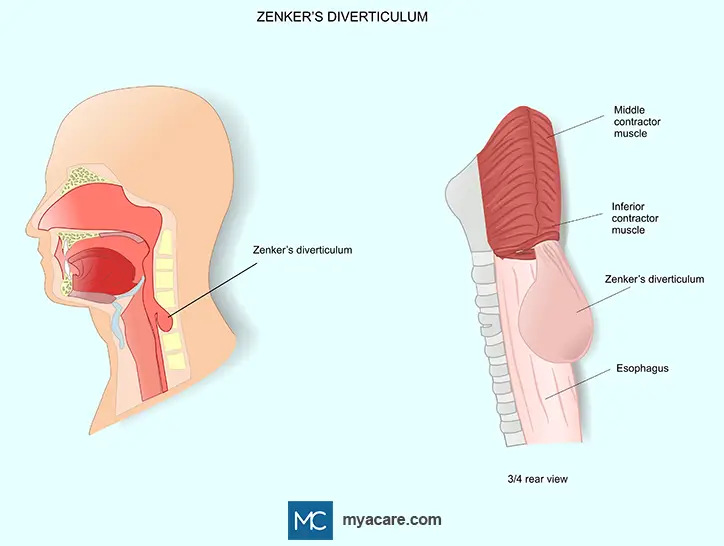
Zenker’s diverticulum is a type of pharyngoesophageal diverticulum. It involves the formation of a pouch that protrudes outward from a weak area in the pharyngeal wall, just above the upper esophageal sphincter. Food and liquids may become trapped in the pouch, leading to choking, aspiration into the lungs, chronic cough, malnutrition, and recurrent respiratory infections.
The pouch forms at the junction between the pharynx and the esophagus, specifically at the back of the throat just above the cricopharyngeus muscle. This area is known as Killian’s triangle, a naturally weak zone between muscle fibers. Because Zenker’s diverticulum lacks a full muscle layer, it is considered a false diverticulum rather than a true one.
Anatomically, the diverticulum arises posteriorly at the top of the esophagus. As it enlarges, it can extend downward and sideways, sometimes becoming large enough to be felt as a neck mass. Its position explains many of the characteristic symptoms, including regurgitation and gurgling sounds in the throat.
Causes of Zenker’s Diverticulum
The primary cause of Zenker’s diverticulum is dysfunction of the cricopharyngeal muscle, which forms the upper esophageal sphincter. Under normal circumstances, this muscle relaxes during swallowing to allow food to pass smoothly into the esophagus. In affected individuals, relaxation is incomplete or delayed.
When the muscle at the top of the food pipe (the cricopharyngeus muscle) fails to relax properly, pressure builds up in the throat (pharynx) during swallowing. Over time, this increased pressure forces the mucosal lining outward through a weak spot in the throat wall, known as Killian’s triangle, forming a pouch. This process explains why Zenker’s diverticulum typically develops slowly over many years.
Food becomes trapped because the pouch sits above the esophagus and empties poorly. Gravity, repeated swallowing, and weak muscle contraction allow food debris and thick mucus to accumulate. This retained material causes irritation, inflammation, and bacterial overgrowth, contributing to bad breath and infection risk.
Excess mucus production is common in Zenker’s diverticulum as the pouch lining responds to chronic irritation.
Risk Factors for Zenker’s Diverticulum
- Advancing age: Most common after 60 due to weakening muscles and reduced sphincter coordination.
- Male gender: Men are affected more often than women.
- Esophageal dysmotility: Abnormal swallowing pressures increase pouch formation risk.
- Neurological conditions: Stroke, Parkinson’s, or other nerve disorders can impair swallowing.
- Previous neck surgery or radiation: May alter anatomy or nerve function, increasing risk.
- Muscle/connective tissue disorders: Reduced pharyngeal strength or elasticity makes herniation more likely.
These factors explain why Zenker’s diverticulum typically develops in older adults and is linked to impaired swallowing mechanics.
Zenker’s Diverticulum Symptoms
Zenker’s diverticulum develops gradually, and symptoms worsen as the pouch enlarges. They are mainly due to impaired swallowing and retention of food and secretions:
- Difficulty swallowing (dysphagia): Most common early symptom. Initially affects solids, later liquids. Patients often feel food sticking in the throat and may need repeated swallows to clear it.
- Regurgitation of undigested food: Trapped food can return to the mouth hours later, usually without nausea. It often occurs when bending or lying down and may have an unpleasant taste or odor.
- Chronic cough or choking: Small amounts of food, saliva, or mucus can enter the airway, causing coughing during meals or at night, and occasional choking episodes.
- Bad breath (halitosis): Bacterial breakdown of retained food causes persistent odor, often noticeable despite good oral hygiene.
- Neck lump or globus sensation: A visible neck swelling that may enlarge after eating and reduce after regurgitation, or a persistent feeling of fullness in the throat (globus sensation), even when not swallowing.
- Hoarseness (dysphonia): Voice changes from nerve irritation or chronic aspiration, causing a hoarse or weak voice.
- Boyce sign: Gurgling sound or sensation when pressing the neck over the diverticulum, caused by movement of air or fluid.
Complications of Zenker’s Diverticulum
Zenker’s diverticulum, if left untreated, can lead to several significant complications:
- Aspiration pneumonia: One of the most serious complications is aspiration pneumonia. Food, saliva, or mucus trapped in the diverticular pouch can spill into the airway, introducing bacteria and triggering lung infection. Repeated aspiration events increase the risk of chronic respiratory illness, hospitalization, and even death in older adults or those with compromised lung function.
- Breathing problems: Large pouches or repeated aspiration can cause nighttime choking, coughing, or shortness of breath; very large diverticula may compress the airway.
- Malnutrition: Difficulty swallowing, fear of choking, and regurgitation often lead patients to reduce their food intake. Over time, this can result in malnutrition, unintentional weight loss, and vitamin deficiencies. In severe cases, dehydration may also occur, particularly if liquids are difficult to swallow.
- Rare risk of cancer: Although uncommon, Zenker’s diverticulum carries a small risk of developing squamous cell carcinoma within the pouch. Chronic irritation, inflammation, and long-term retention of food and secretions are thought to contribute to this malignant transformation. This makes long-term monitoring important, especially in older adults or patients with longstanding diverticula.
- Potentially life-threatening: While Zenker’s diverticulum itself is benign, its complications can be life-threatening. Aspiration pneumonia is the leading cause of mortality associated with the condition. Other potentially life-threatening outcomes can also result from diverticular perforation, malnutrition/dehydration from chronic dysphagia, and rarely, bleeding from an ulcerated pouch and squamous cell carcinoma arising within the pouch. Mortality is particularly high in elderly patients or those with comorbidities. Early diagnosis and management significantly reduce the risk of severe outcomes.
Zenker’s Diverticulum Diagnosis
Accurate diagnosis of Zenker’s diverticulum is essential to prevent complications and guide appropriate treatment. Multiple diagnostic tools and evaluations are used:
-
Zenker’s Diverticulum Classification
Diverticula are commonly classified by size:
-
- Small: less than 2 centimeters
- Medium: 2–4 centimeters
- Large: greater than 4 centimeters
Size impacts symptoms, treatment choice, and risk of complications.
-
Barium Swallow Study (Gold Standard)
The barium swallow is the most reliable test for detecting Zenker’s diverticulum. The patient swallows a radiopaque liquid while X-rays capture the flow through the pharynx and esophagus. This imaging reveals the size, location, and emptying characteristics of the pouch and helps differentiate it from other structural abnormalities.
-
Endoscopy
Endoscopy is used to directly visualize the esophagus and pharynx. It is particularly useful for ruling out malignancy or assessing the mucosal lining. Endoscopy must be done carefully in Zenker’s diverticulum because the endoscope can inadvertently enter the pouch, creating a risk of perforation.
-
Esophageal Manometry
Esophageal manometry is not routinely performed to diagnose Zenker’s diverticulum, but it may be considered when a coexisting esophageal motility disorder is suspected.
-
Coexistence With Achalasia
When both conditions are clinically significant, swallowing difficulties may reflect dysfunction at both the upper and lower esophageal sphincters. Treatment should therefore be individualized and may require management of both the Zenker’s diverticulum and achalasia.
Differential Diagnosis
Other conditions that can mimic Zenker’s diverticulum must be considered:
Repeated exposure to gastric contents irritates the esophagus and may cause symptoms such as difficulty in swallowing, heartburn, or sensation of a lump in the throat.
-
Achalasia
Lower esophageal muscle fails to relax, preventing food from passing easily into the stomach and causing regurgitation.
-
Esophagitis
Inflammation of the esophageal lining often caused by infection, medication, or radiation therapy; can lead to painful or difficult swallowing.
Chronic acid reflux damages the lower esophageal lining, causing it to change and potentially leading to swallowing difficulties or heartburn.
-
Esophageal squamous cell carcinoma
Originates in the squamous cells lining the esophagus and causes progressive difficulty swallowing.
-
Killian-Jamieson diverticulum
Arises lateral to the cricopharyngeus muscle and is generally smaller and less symptomatic than Zenker’s.
-
Epiphrenic diverticulum
Occurs near the lower esophagus, often associated with motility disorders.
-
Cricopharyngeal bar
A prominent upper esophageal sphincter seen on imaging, which may cause mild dysphagia but does not form a true pouch.
-
Hiatal hernia
Part of the stomach protrudes through the diaphragm; can cause reflux but is anatomically distinct.
-
Plummer-Vinson Syndrome (PVS)
Characterized by esophageal webs and iron deficiency anemia; may cause dysphagia but not diverticulum formation.
Careful differentiation between these conditions is essential because management strategies vary widely depending on the exact diagnosis. Barium swallow combined with endoscopic evaluation is usually sufficient to confirm Zenker’s diverticulum and exclude other possibilities.
Treatment Options for Zenker’s Diverticulum
Management of Zenker’s diverticulum depends on the size of the pouch, severity of symptoms, patient age, and overall health. Treatment ranges from observation for small, asymptomatic pouches to minimally invasive endoscopic procedures and open surgical repair for larger or symptomatic diverticula. Treatment is designed to ease symptoms, minimize complications, and help patients swallow safely.
Observation – Small, Asymptomatic Cases
For small diverticula that do not cause significant swallowing difficulty, regurgitation, or aspiration risk, a conservative approach may be appropriate. Observation includes:
- Regular monitoring with follow-up visits and imaging as needed
- Dietary adjustments to reduce stress on the upper esophageal sphincter
- Education on recognizing early signs of worsening symptoms or complications
Observation is appropriate only for truly asymptomatic or minimally symptomatic patients because diverticula tend to enlarge over time. Periodic reassessment can help determine whether intervention is required.
Non-Surgical / Minimally Invasive Treatments
Minimally invasive procedures are often preferred for older adults or patients with comorbidities who may face greater risks with open surgery. These approaches aim to divide the cricopharyngeal muscle (myotomy) and allow the pouch to empty directly into the esophagus.
Endoscopic stapling
- A staple line divides the common wall between the diverticulum and esophagus, creating a single channel for food passage.
- Benefits: Short procedure time, rapid recovery, minimal external scarring.
- Limitations: Recurrence is possible, particularly with very large pouches.
Rigid and flexible endoscopic diverticulotomy
- A flexible or rigid endoscope is used to cut the septum between the esophagus and the pouch.
- Benefits: Flexible techniques are often better tolerated in elderly patients.
- Limitations: Risks include bleeding, perforation, and infection.
Peroral endoscopic myotomy (POEM)
- Originally developed for achalasia, POEM can be adapted for Zenker’s diverticulum.
- It involves creating a submucosal tunnel and cutting the cricopharyngeal muscle from within.
- Benefits: Minimally invasive with reduced hospital stay.
- Limitations: Perforation remains a possible complication if the mucosal incision does not seal properly.
Dohlman procedure
- The Dohlman procedure was an early endoscopic treatment for Zenker’s diverticulum that established the principle of dividing the diverticular septum and cricopharyngeal muscle, forming the basis for later endoscopic techniques.
- Benefits: Provides symptom relief through a direct approach to septum division.
- Limitations: Higher risks of bleeding and perforation, leading to its replacement by modern adaptations.
Laser surgery
- A laser is used to divide the septum between the esophagus and pouch.
- Benefits: Precise cutting, minimal bleeding.
- Limitations: Requires specialized equipment and skill; risk of recurrence remains.
Minimally invasive treatments generally have shorter recovery times compared with open surgery and are associated with lower postoperative morbidity, but not all patients are suitable candidates, particularly those with very large diverticula or extensive scar tissue.
When Is Open Surgery Considered?
Open surgical approaches are reserved for large diverticula, failed endoscopic treatment, or complicated cases.
Diverticulectomy and diverticulopexy
- Diverticulectomy: Surgical removal of the pouch.
- Diverticulopexy: The pouch is suspended and stitched to prevent food accumulation, often combined with cricopharyngeal myotomy.
Indications for surgery
- Large diverticula causing significant dysphagia
- Recurrent aspiration or chronic infection
- Suspicion of malignancy within the pouch
How long does Zenker's diverticulum surgery take?
Surgery is performed under general anesthesia, with the typical duration ranging from one to two hours, depending on the complexity and size of the diverticulum.
Risks
- Bleeding and infection
- Injury to the recurrent laryngeal nerve, potentially causing hoarseness or vocal cord paralysis
- Leakage from the surgical site, which may require additional intervention
- Rare cardiopulmonary complications, particularly in older adults
Recovery and Outcomes
Expected Symptom Improvement
- Most patients experience significant improvement in swallowing, reduction in regurgitation, and resolution of aspiration risk.
- Endoscopic procedures often provide immediate relief of dysphagia.
Recovery Time for Different Treatments
- Endoscopic procedures: 1–3 days for most activities; diet may advance rapidly from liquids to solids.
- Open surgery: 2–6 weeks for full recovery; gradual diet progression is recommended.
Surgical Success Rate
- Success rates are high, often exceeding 90% for both endoscopic and open approaches.
- Although recurrence rates are generally low, the condition can recur, especially after endoscopic procedures, necessitating repeat intervention in some patients.
Postoperative complications
- Breathing problems may occur temporarily due to swelling or airway irritation.
- Voice changes may appear if nearby nerves are affected.
Speech therapy
- Postoperative rehabilitation may include swallowing therapy to restore coordination and prevent aspiration.
- Speech-language pathologists can provide exercises to improve strength, timing, and safety of swallowing.
Living With Zenker’s Diverticulum
Dietary adjustments play an important role in symptom management both before and after treatment.
Eating and Swallowing Tips
- Eat at a slow pace and chew food well to help keep it from entering the pouch.
- Take small bites and sip fluids during meals to help clear food efficiently.
- Maintain an upright posture while eating and remain upright for 30–60 minutes afterward.
Foods to Avoid
- Dry, sticky, or coarse foods that may lodge in the pouch (e.g., tough meats, crackers, or bread).
- Large boluses that require forceful swallowing.
Diet After Zenker’s Diverticulum Surgery
- Begin with liquids immediately postoperatively.
- Gradually advance to soft foods, then regular textures over 1–2 weeks, depending on surgical approach and healing.
- Follow dietary instructions provided by the surgical and speech therapy teams to minimize complications and support recovery.
Proper adherence to dietary recommendations, combined with surgical or endoscopic treatment, significantly improves quality of life, reduces aspiration risk, and ensures long-term success in managing Zenker’s diverticulum.
Frequently Asked Questions
-
Can Zenker’s diverticulum go away on its own?
No. Zenker’s diverticulum generally does not resolve without treatment and usually enlarges over time.
-
Does Zenker’s diverticulum cause snoring?
Zenker’s diverticulum is not a well-established cause of snoring. However, a larger pouch may retain food that regurgitates into the throat, potentially causing nighttime coughing or choking.
-
Can Zenker’s diverticulum be hereditary?
Zenker’s diverticulum is usually sporadic, and a genetic cause has not been established. However, rare cases occurring within the same family have been reported.
-
Do pharyngeal pouches get bigger?
Yes. The pouch generally enlarges gradually if untreated, worsening symptoms over time.
To search for the best ENT Doctors and Healthcare Providers worldwide, please use the Mya Care search engine.
To search for the best Gastroenterology Doctors and Healthcare Providers worldwide, please use the Mya Care Search engine.
To search for the best doctors and healthcare providers worldwide, please use the Mya Care search engine.
The Mya Care Editorial Team comprises medical doctors and qualified professionals with a background in healthcare, dedicated to delivering trustworthy, evidence-based health content.
Our team draws on authoritative sources, including systematic reviews published in top-tier medical journals, the latest academic and professional books by renowned experts, and official guidelines from authoritative global health organizations. This rigorous process ensures every article reflects current medical standards and is regularly updated to include the latest healthcare insights.

Dr. Sony Sherpa completed her MBBS at Guangzhou Medical University, China. She is a resident doctor, researcher, and medical writer who believes in the importance of accessible, quality healthcare for everyone. Her work in the healthcare field is focused on improving the well-being of individuals and communities, ensuring they receive the necessary care and support for a healthy and fulfilling life.

Dr. Dora Matis is a licensed medical doctor based in Germany, specializing in clinical psychiatry and psychotherapy. With a strong academic background and international training, she brings expertise in evidence-based medical practice. In addition to her clinical work, Dr. Matis has been involved in translational research, helping bridge the gap between scientific discovery and patient care.
References:
Featured Blogs

Medically Induced Coma: What It Is, How It Works & Recovery