
What Is Sarcoidosis? Symptoms, Causes, and Treatment
Medically Reviewed by Dr. Sony Sherpa, (MBBS) - October 06, 2026
Fact Checked and Edited by Dr. Asif Baliyan, MD - October 06, 2026
Key Takeaways
- Sarcoidosis is an inflammatory disease marked by clusters of immune cells that most often affect the lungs and lymph nodes but can involve many organs.
- Its exact cause is unknown, although immune dysregulation, genetic susceptibility, infections, and environmental exposures are discussed as possible contributors.
- Symptoms vary by organ involvement and may include respiratory, skin, eye, cardiac, neurological, musculoskeletal, and systemic problems.
- Diagnosis may involve imaging, laboratory and functional tests, and biopsy, while treatment ranges from observation to corticosteroids, steroid-sparing drugs, biologic therapy, and supportive care.
- Prognosis varies widely; many cases resolve, while chronic disease can lead to fibrosis or serious cardiac, neurological, and other complications.
Introduction
Sarcoidosis is an inflammatory condition that can affect different parts of the body and varies considerably from person to person. Some people experience mild or temporary symptoms, while others develop chronic inflammation that can lead to organ damage.
Sarcoidosis occurs worldwide, though its frequency differs across populations. In England, studies show a rising prevalence, increasing from roughly 167 to 230 per 100,000 individuals over the past two decades.
In the United States, it is significantly more common among African Americans, with an incidence of approximately 35.5 per 100,000, compared with around 10 per 100,000 among White Americans.
Globally, rates vary widely: Northern European countries report some of the highest incidence, while certain regions in Asia show much lower rates.
Several public figures have spoken openly about living with sarcoidosis, helping raise awareness and reduce stigma around the condition. Actress Tisha Campbell has publicly discussed her journey with sarcoidosis, including periods of remission and the importance of managing stress in maintaining her health.
The late comedian Bernie Mac lived with sarcoidosis, and his experience inspired the creation of the Bernie Mac Foundation, which promotes research and education about the disease. Actress and writer Karen Duffy was diagnosed with neurosarcoidosis, a form affecting the nervous system, and has openly shared her experience living with chronic pain, helping others understand the challenges of the condition.
What Is Sarcoidosis?
Sarcoidosis is an inflammatory disease in which the body’s immune system becomes overactive and leads to the formation of tiny clumps of immune cells called granulomas. These granulomas can develop in almost any organ, but they most commonly affect the lungs, lymph nodes, skin, and eyes. The condition varies widely between individuals; some people experience mild, temporary illness, while others develop chronic inflammation that leads to long-term organ damage.
Immune Dysregulation: What Goes Wrong
Although the exact cause of sarcoidosis remains unknown, the disease is thought to stem from a dysregulated immune response. Instead of responding normally to a trigger, which may be environmental, infectious, or unknown, the immune system becomes excessively activated.
Key immune processes involved include:
-
Overactivation of T-helper (CD4+) cells:
These cells normally coordinate immune responses, but in sarcoidosis they become hyper-responsive. They accumulate at the site of inflammation and recruit more immune cells than necessary.
-
Activation of macrophages:
Macrophages, another major immune cell type, join the CD4+ cells. They release highly active inflammatory substances and transform into “epithelioid cells,” which are essential components of granulomas.
-
Cytokine release:
Activated CD4+ cells and macrophages produce a cascade of inflammatory cytokines—including TNF-α, IL-2, and interferon-γ. These chemical messengers amplify inflammation, sustain the immune response, and drive the formation of granulomas.
Formation of Non-Caseating Granulomas
The hallmark of sarcoidosis is the presence of non-caseating granulomas – tightly organized clusters of immune cells that do not have the cheese-like center seen in infections such as tuberculosis.
These granulomas are the body’s attempt to “wall off” what it perceives as a threat. But in sarcoidosis, the threat may be unclear or no longer present, so granulomas persist unnecessarily.
How Granulomas Cause Organ Damage
Granulomas themselves are not inherently harmful, but their presence in large numbers disrupts normal tissue structure. Over time, this leads to:
- Ongoing inflammation in the affected organ
- Fibrosis (scarring) as the body attempts to repair the chronic inflammation
- Loss of normal organ function when scarring becomes extensive
For example, granulomas in the lungs can stiffen lung tissue and reduce breathing capacity, while granulomas in the heart or nervous system can cause more serious, sometimes life-threatening complications.
Causes and Risk Factors of Sarcoidosis
The exact cause of sarcoidosis remains unknown. Most researchers believe it develops from an overactive immune response in someone who is genetically susceptible, triggered by certain environmental or infectious exposures.
Proposed Triggers
While no single cause has been proven, several potential triggers have been linked to sarcoidosis:
Infectious agents
Researchers have explored whether certain microbes may stimulate the abnormal immune reaction. These include:
- Mycobacterium species
- Propionibacterium acnes
- Various viruses (e.g., herpes viruses)
These infections are not considered contagious causes; rather, they may act as immune triggers.
Environmental exposures
Environmental or occupational exposures may also play a role in initiating granuloma formation:
- Dusts and metals (e.g., beryllium, aluminum)
- Mold or organic dust
- Pesticides
- Firefighting environments
- Agricultural or industrial chemicals
These exposures do not cause sarcoidosis on their own, but they may be contributing factors in people with genetic vulnerability.
Genetic susceptibility
Genetics appear to influence:
- Who develops sarcoidosis
- Severity of the disease
- Which organs are affected
Inherited variations in immune-regulation genes (e.g., HLA class II genes) have been associated with higher risk.
Risk Factors for Developing Sarcoidosis
Family history
Sarcoidosis is not directly hereditary, but having a close relative (parent, sibling) increases the likelihood of developing the condition. This suggests a genetic predisposition, not a strictly inherited disease.
Age
Sarcoidosis can occur at any age, but it most commonly affects:
- Adults between 20–50 years old
- A smaller second peak occurs in older adults in some populations
Gender
Patterns vary by region, but in many studies:
- Women have a slightly higher risk of developing sarcoidosis
- Women are also more likely to experience chronic or severe disease patterns
Ethnicity
Sarcoidosis rates differ significantly between ethnic groups:
- Higher prevalence and more severe disease in African, African American, Caribbean, and Scandinavian populations
- More chronic and organ-involving forms seen in these groups
- Lower prevalence in Asian populations, though regional clusters exist
Sarcoidosis Symptoms
Sarcoidosis can affect almost any organ. Some people have mild symptoms; others have multi-organ involvement. Many symptoms stem from granuloma-related inflammation, which disrupts normal organ function.
General (Systemic) Symptoms
These are common early signs and reflect widespread inflammation:
- Fatigue (one of the most common and persistent symptoms)
- Fever
- Unintentional weight loss
- Night sweats
- Malaise/feeling generally unwell
- Loss of appetite
- Joint pains or generalized aches
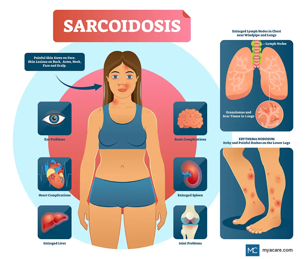
Organ-specific symptoms are discussed below.
Lung Symptoms (Most Commonly Affected Organ)
Up to 90% of patients have lung involvement, known as pulmonary sarcoidosis.
Common lung symptoms include:
- Persistent dry cough
- Shortness of breath (dyspnea)
- Chest discomfort or pain
- Wheezing (less common)
Stages of pulmonary sarcoidosis (chest X-ray classification)
These stages help describe lung involvement – they do not predict severity.
Stage 0:
- Normal chest X-ray
- No visible lung involvement
Stage I:
- Bilateral hilar lymphadenopathy (swollen lymph nodes in the chest)
- Minimal or no lung infiltrates
Stage II:
- Lymphadenopathy plus lung infiltrates
- May show nodules or interstitial changes
Stage III:
- Lung infiltrates without lymphadenopathy
- More inflammation in lung tissue
Stage IV:
- Pulmonary fibrosis
- Scarring leads to reduced lung function
- May show honeycombing and architectural distortion
Calcified granulomas in the lungs
- Granulomas sometimes heal and calcify, appearing as small white spots on imaging.
- Calcification usually indicates past or chronic inflammation, not active disease.
- These are sometimes confused with TB calcifications, but sarcoid granulomas are non-caseating.
Lymph Node Symptoms
Lymph node swelling is very common.
Symptoms & findings may include:
- Enlarged lymph nodes in:
- Chest (hilar/mediastinal)
- Neck
- Underarms
- Groin
- Usually non-tender
- May be found incidentally on imaging
Skin (Cutaneous Sarcoidosis) Symptoms
Skin involvement appears in about 20–35% of patients.
Common skin manifestations include:
-
Erythema nodosum:
-
Tender red nodules, usually on the shins
-
Often associated with acute sarcoidosis
-
-
Lupus pernio:
-
Chronic, violaceous (purple) plaques
-
Typically on nose, cheeks, lips, and ears
-
Indicates more severe systemic disease
-
-
Maculopapular lesions:
-
Small bumps or patches, often on the face, arms, or trunk
-
-
Scar infiltration:
-
Old scars become raised, swollen, or discolored
-
-
Hypo- or hyperpigmented papules or plaques:
-
Skin lesions that appear lighter or darker than the surrounding skin
-
Eye (Ocular Sarcoidosis) Symptoms
Approximately 20–30% of patients have eye involvement.
Symptoms include:
- Redness
- Eye pain
- Blurry vision
- Dry eyes
- Light sensitivity
Common ocular conditions
- Uveitis
- Conjunctival granulomas
- Lacrimal gland involvement, leading to dry eyes
Liver and Spleen Symptoms
Although often asymptomatic, granulomas are frequently detected on imaging or biopsy.
Liver (hepatic sarcoidosis)
- Elevated liver enzymes
- Right upper abdominal discomfort
- Enlarged liver (hepatomegaly)
- Rarely, portal hypertension or cirrhosis
Spleen
- Enlarged spleen (splenomegaly)
- Low blood cell counts (if hypersplenism develops)
Heart (Cardiac Sarcoidosis) Symptoms
Potentially serious and sometimes life-threatening.
Symptoms may involve:
- Palpitations
- Shortness of breath
- Dizziness or fainting (syncope)
- Chest pain
- Fatigue due to heart block or arrhythmias
Possible complications
- Heart block
- Ventricular arrhythmias
- Heart failure
- Sudden cardiac death
Nervous System (Neurosarcoidosis) Symptoms
Affects 5–10% of patients, involving the brain, spinal cord, or nerves.
Symptoms may include:
- Facial nerve palsy (sudden facial drooping)
- Peripheral neuropathy (numbness, tingling, burning)
- Seizures
- Headaches
- Aseptic Meningitis (inflammation of the meninges around the brain)
- Lower extremity weakness
- Visual changes (optic nerve involvement)
- Hormonal issues (pituitary involvement)
Symptoms of the Joints and Muscles
Sarcoidosis can cause musculoskeletal symptoms.
Joint symptoms
- Arthralgia (joint pain)
- Joint swelling (ankles most common)
- Pain in shoulders, knees, wrists
- Granulomatous inflammation and periarthritis can cause shoulder discomfort.
Muscle symptoms
- Muscle weakness
- Tender nodules
- Granulomatous myopathy (rare)
Mouth and Upper Airway Symptoms
Though less common, oral involvement can occur.
Symptoms include:
- Granulomas on lips, gums, or oral mucosa
- Swelling of salivary glands
- Dry mouth
- Hoarseness (if the larynx is involved)
- Persistent oral ulcers (uncommon)
Potential Complications
Sarcoidosis can be mild for many people, but in others it may lead to long-term complications due to persistent inflammation, granuloma formation, or organ damage. The likelihood of complications depends on which organs are involved and how active the disease remains over time.
Pulmonary Fibrosis and Respiratory Failure
Chronic lung inflammation can lead to fibrosis, a form of permanent scarring where healthy lung tissue is replaced with stiff, non-functional tissue. As fibrosis progresses, it reduces lung capacity and gas exchange.
Severe cases may result in:
- Persistent shortness of breath
- Low oxygen levels
- Exercise intolerance
- Respiratory failure, requiring long-term oxygen therapy or, rarely, lung transplantation
Cardiac Arrhythmias and Heart Block
Cardiac sarcoidosis can disrupt the heart’s electrical conduction system. When granulomas form in heart tissue, they may interrupt normal signaling pathways.
Possible complications include:
- Arrhythmias (irregular heartbeats)
- Complete or partial heart block
- Ventricular tachycardia or fibrillation (potentially life-threatening)
- Sudden cardiac death in severe, untreated cases
Early detection and management are essential, especially if symptoms like palpitations, dizziness, or fainting occur.
Vision Loss Due to Chronic Uveitis
Ocular sarcoidosis can lead to chronic uveitis, a long-lasting inflammation inside the eye. Without timely treatment, persistent inflammation may cause:
- Glaucoma
- Cataracts
- Retinal damage
- Optic nerve involvement
These complications can result in partial or permanent vision loss, making regular ophthalmologic evaluations vital for patients with eye symptoms.
Hypercalcemia-Related Kidney Problems
Sarcoidosis can raise calcium levels in the blood (hypercalcemia) or urine (hypercalciuria) because activated granulomas increase vitamin D production.
This can lead to:
- Kidney stones
- Nephrocalcinosis (calcium deposits in kidney tissue)
- Gradual decline in kidney function
Maintaining hydration and monitoring calcium levels are essential in patients prone to these issues.
Chronic Fatigue Syndrome–Like Symptoms
Even when sarcoidosis is otherwise stable, many patients experience severe and persistent fatigue. This can resemble chronic fatigue syndrome and may persist for years.
Fatigue often stems from:
- Ongoing low-grade inflammation
- Sleep disturbances
- Medication effects
- Chronic pain
- Autonomic dysfunction in some cases
Fatigue is one of the most disabling long-term complications and significantly affects quality of life.
Diagnosis
Diagnosing sarcoidosis can be challenging because no single test confirms it. Instead, doctors rely on insights from clinical evaluation, imaging, laboratory tests, and tissue biopsy to rule out other causes of granulomatous disease.
Clinical Evaluation
Diagnosis begins with a detailed medical history and physical examination. Clinicians assess:
- Duration and pattern of symptoms (fatigue, cough, weight loss, fever)
- Organ-specific findings
- Occupational or environmental exposures
- Family history
- Signs of organ involvement (e.g., skin lesions, enlarged lymph nodes, arrhythmias, eye redness)
Imaging
Chest X-ray
Often the first imaging test used. It helps identify:
- Bilateral hilar lymphadenopathy
- Lung infiltrates
- Patterns used to classify stages of pulmonary sarcoidosis, which are outlined above
High-resolution CT (HRCT)
More detailed imaging used to detect:
- Granulomas
- Fibrosis
- Airway distortion
- Calcified granulomas in the lungs
HRCT helps distinguish sarcoidosis from infections or malignancy.
Nuclear imaging
Useful when assessing heart or whole-body involvement:
- PET scan
- Gallium scan
These investigations can help detect areas of increased metabolic activity or active inflammation and identify biopsy sites
Laboratory Tests
Common lab abnormalities include:
- Elevated ACE (angiotensin-converting enzyme)
- Hypercalcemia or hypercalciuria due to increased vitamin D activation
- Elevated ESR and CRP
- Abnormal liver enzymes (ALP, AST/ALT) if the liver is affected
- Lymphopenia
- Elevated soluble IL-2 receptor levels (in some centers)
None of these tests alone confirm sarcoidosis, but they support the diagnosis.
Functional Tests
Depending on organ involvement:
Pulmonary function tests (PFTs)
May show:
- Restrictive pattern
- Reduced DLCO (gas exchange).
Slit-lamp examination
For ocular sarcoidosis:
- Detects uveitis
- Retinal vasculitis
- Conjunctival granulomas.
Cardiac evaluation
- ECG, echocardiogram
- Cardiac MRI
- PET scan.
Procedures for Tissue Diagnosis (Biopsy)
Biopsy is the gold standard, showing non-caseating granulomas.
Common biopsy sites:
- Skin lesions
- Peripheral lymph nodes
- Transbronchial lung biopsy
- EBUS-TBNA (Endobronchial Ultrasound-Guided Needle Aspiration)
- Bronchoscopic transbronchial biopsy
- Mediastinoscopy (for deeper lymph nodes)
- Liver biopsy (if abnormal liver tests or organ involvement).
Differential Diagnosis
Sarcoidosis must be distinguished from other causes of granulomatous disease:
An important differential, especially in TB endemic areas. TB is more likely to demonstrate caseating/necrotizing granulomas, acid-fast bacilli, positive cultures or molecular tests, although non-necrotizing granulomas can be seen. A tuberculin skin test (TST/ purified protein derivative [PPD]) or interferon-gamma release assay can help identify TB infection but cannot distinguish active TB from sarcoidosis by itself. When TB is suspected, microbiological testing is important.
-
Fungal infections (eg histoplasmosis):
Can cause granulomatous inflammation and hilar/mediastinal lymphadenopathy mimicking sarcoidosis. Diagnosis may be aided by fungal stains and cultures, antigen testing, and epidemiological exposure history.
-
Lymphoma:
May present with lymphadenopathy, constitutional symptoms, and pulmonary findings similar to sarcoidosis. Asymmetric or bulky lymph node enlargement, extranodal disease, and malignant cells on tissue sampling favor lymphoma.
-
Amyloidosis:
Can rarely cause pulmonary nodules, interstitial disease, or lymphadenopathy. Amyloidosis is diagnosed by demonstrating amyloid deposition, usually with Congo red staining and characteristic birefringence (an apple-green appearance under polarized light), as opposed to sarcoidosis.
-
Lupus and other autoimmune diseases:
Systemic autoimmune diseases may have constitutional, pulmonary, joint, skin, or other manifestations that overlap with sarcoidosis. They are distinguished by the overall clinical picture and by autoantibodies.
-
Hypersensitivity pneumonitis:
Radiologically and histologically, it may resemble pulmonary sarcoidosis. History of exposure to mold, birds, agricultural dust, or other inhaled antigens predisposes to hypersensitivity pneumonitis, with centrilobular nodules, air trapping, and poorly formed airway-centered granulomas.
-
Granulomatosis with polyangiitis (GPA; formerly Wegener granulomatosis):
GPA may present with upper-airway/sinus disease, pulmonary lesions with cavities, and renal involvement. Testing for ANCA (antineutrophil cytoplasmic antibodies; a blood test to identify antibodies associated with GPA) and a biopsy demonstrating necrotizing vasculitis may help confirm the diagnosis.
-
Chronic beryllium disease:
It can be very difficult to differentiate from sarcoidosis as both may show non-necrotizing granulomas and similar pulmonary imaging findings. A detailed occupational history and beryllium lymphocyte proliferation testing (BeLPT) are particularly important in the setting of beryllium exposure.
-
Cardiac sarcoidosis vs. myocarditis:
Both can present with arrhythmias, conduction abnormalities, inflammation of the heart muscle, and ventricular dysfunction. Cardiac MRI and FDG-PET may show patterns of inflammation or scarring, but interpretation requires correlation with the clinical context.
-
Disorders related to rheumatoid arthritis and other inflammatory arthritides:
May have joint, pulmonary, and nodular manifestations that overlap with sarcoidosis. They can be differentiated by clinical phenotype, serology, imaging, and biopsy.
-
Sarcoidosis vs. mesothelioma:
In patients with pleural abnormalities, especially in patients with a history of asbestos exposure, mesothelioma should be on the differential. Pleural thickening, nodularity, effusion, and biopsy evidence of malignant cells favor mesothelioma over sarcoidosis.
Importantly, the finding of non-caseating granulomas is not by itself diagnostic for sarcoidosis. The diagnosis should be made after exclusion of infectious organisms and other granulomatous disorders.
Treatment
Treatment depends on organ involvement, severity, and risk of long-term damage. Many cases resolve spontaneously and require only monitoring.
Observation
Some people do not need treatment, especially those with:
- Mild symptoms
- Asymptomatic Stage I lung disease
- Löfgren’s syndrome (fever, erythema nodosum, ankle arthritis, and lymphadenopathy), which often resolves on its own
Regular follow-up ensures early detection of progression.
Medical Treatment
First-line: Corticosteroids
- Prednisone is the primary treatment
- Reduces inflammation and granuloma formation
- Typical uses: lungs, eyes, skin, joints, heart, and nervous system
Side effects to monitor: Weight gain, mood changes, diabetes, hypertension, osteoporosis, and fluid retention.
Second-line: Steroid-sparing agents
Used when steroids are ineffective, cause side effects, or long-term therapy is required.
Common options:
- Methotrexate
- Azathioprine
- Mycophenolate mofetil
- Hydroxychloroquine (especially for skin and calcium issues)
Refractory cases
For severe or treatment-resistant disease:
- Anti–TNF-alpha therapies (e.g., infliximab, adalimumab)
- Cyclophosphamide (severe neurosarcoidosis)
- Rituximab (rare cases)
Supportive therapies
- NSAIDs for joint or musculoskeletal pain
- Physical therapy to improve mobility and function
- Pulmonary rehabilitation for breathlessness and fatigue
Cardiac devices
Patients with cardiac sarcoidosis may require:
- Implanted pacemaker
- Implantable cardioverter-defibrillator (ICD)
These prevent fatal arrhythmias or heart block.
Complications/Side Effects of Treatment
Long-term steroid or immunosuppressant use may lead to:
- Diabetes
- Hypertension
- Osteoporosis
- Increased infection risk
- Liver toxicity
- Gastrointestinal issues
Lifestyle Changes and Management
Lifestyle modifications can help control symptoms, prevent complications, and support medical treatment.
How to Reduce the Risk of Sarcoidosis Progression
- Do not smoke and do not be around tobacco smoke
- When possible, stay away from workplace or environmental irritants (for example, dust, chemicals, molds)
- Keep a healthy weight and diet
- Be physically active according to your tolerance level
- Take medications as prescribed and attend regular follow-up appointments
- Monitor lung function and other organ systems as per the affected organs
Diet and Medication Considerations
The following should be considered, especially in hypercalcemic or hypercalciuric patients:
- If you have high calcium levels, watch vitamin D–fortified foods and drinks.
- Limit the use of antacids containing calcium.
- Dairy products need not be routinely removed. Dietary calcium restriction should be individualized based on serum and urinary calcium levels.
Long-term corticosteroids should be used in conjunction with a well-rounded diet containing adequate calcium and vitamin D (not automatic calcium restriction, as corticosteroids increase the risk of osteoporosis).
What supplements should I avoid with sarcoidosis?
Granulomas can cause excessive vitamin D activation, raising calcium levels.
- Do not take vitamin D supplements unless under the advice and supervision of your health care provider.
- Do not take calcium supplements unless your doctor tells you to, especially if you have too much calcium in your blood or urine.
- Activated macrophages in sarcoid granulomas may secrete extrarenal 1α-hydroxylase, leading to increased conversion of 25-hydroxyvitamin D to the active form 1,25-dihydroxyvitamin D (calcitriol). It may increase intestinal absorption of calcium and lead to hypercalcemia and hypercalciuria.
Importantly, vitamin D is not universally contraindicated in sarcoidosis. The American Thoracic Society recommends measuring both 25-hydroxyvitamin D and 1,25-dihydroxyvitamin D before starting replacement therapy when supplementation is being considered.
Natural Ways to Support Sarcoidosis Management
These can be used in addition to medical treatment, but not as a replacement for prescribed therapy:
- Eat a healthy, balanced diet that is rich in nutrients instead of a specific “anti-inflammatory” diet.
- Perform suitable exercise, whether it be walking, yoga, or formalized exercise, per individual tolerance.
- Get enough sleep on a regular basis.
- Try stress reduction techniques such as mindfulness, meditation, and breathing exercises.
- Turmeric/curcumin and omega-3 fatty acids: Evidence for a specific benefit for sarcoidosis is currently limited, and these should not be regarded as established therapies.
- Talk to your doctor before taking supplements, as vitamin D and calcium metabolism may be abnormal in sarcoidosis.
Exercise, pulmonary rehabilitation, and mindfulness-based interventions have evidence for improving fatigue and quality of life in selected patients with sarcoidosis.
Home Remedies
Supportive measures can help relieve symptoms, but should not take the place of medical treatment:
- Hot packs for joint or musculoskeletal pain
- Humidifier for a dry throat or airway
- Adequate hydration, particularly in patients with hypercalciuria, when medically appropriate
- Sun protection, particularly in patients with hypercalcemia or hypercalciuria
Prognosis and Outlook
Sarcoidosis has a highly variable course.
Can Sarcoidosis Go Away?
Yes. In many people, especially those with Stage I or II lung disease or Löfgren’s syndrome, sarcoidosis can resolve spontaneously.
Overall Prognosis
The majority of patients have a good prognosis, while about one-third develop chronic disease and a smaller group may experience severe complications.
Worse prognosis is associated with:
- Cardiac involvement
- Neurological involvement
- Advanced pulmonary fibrosis
Sarcoidosis Life Expectancy
Most individuals live normal lifespans. Mortality usually relates to:
- Advanced lung fibrosis
- Cardiac arrhythmias
- Neurosarcoidosis
Latest Research and Advancements
Efzofitimod
New studies are exploring efzofitimod, a biologic therapy targeting immune pathways involved in granuloma formation. Early results show:
- Reduced steroid requirements
- Improved lung function
- Lower inflammation markers
Breakthrough Protein Research
Recent discoveries about proteins involved in pulmonary sarcoidosis may lead to:
- New diagnostic biomarkers
- More precise targeted therapies
- Better prediction of disease severity
These emerging treatments offer hope for patients with refractory or advanced disease.
Frequently Asked Questions
Is sarcoidosis cancer?
No. Sarcoidosis is not cancer. However, enlarged lymph nodes can mimic cancer on scans.
Is sarcoidosis an autoimmune disease?
It is considered an immune-mediated disease, with features of both autoimmune and inflammatory disorders.
Are there wearable devices that help monitor sarcoidosis symptoms?
Yes. Smartwatches and wearable monitors can track:
- Heart rate
- Oxygen saturation
- Physical activity
- Sleep
- Arrhythmia alerts (in some devices)
Monitoring these aspects can be useful for cardiac or pulmonary sarcoidosis.
Does sarcoidosis affect teeth?
Indirectly, yes. Steroids may affect gum health, and granulomas can rarely affect salivary glands, causing dry mouth and dental issues.
Does sarcoidosis affect tattoos?
Yes. Sarcoidosis can cause granulomas within old or new tattoos, leading to raised, red, or painful areas.
To search for the best Pulmonary and Respiratory Medicine Doctors and Healthcare Providers in Germany, India, Malaysia, the UAE and the USA please use the Mya Care Search engine.
To search for the best doctors and healthcare providers worldwide, please use the Mya Care search engine.
The Mya Care Editorial Team comprises medical doctors and qualified professionals with a background in healthcare, dedicated to delivering trustworthy, evidence-based health content.
Our team draws on authoritative sources, including systematic reviews published in top-tier medical journals, the latest academic and professional books by renowned experts, and official guidelines from authoritative global health organizations. This rigorous process ensures every article reflects current medical standards and is regularly updated to include the latest healthcare insights.
Dr. Sony Sherpa completed her MBBS at Guangzhou Medical University, China. She is a resident doctor, researcher, and medical writer who believes in the importance of accessible, quality healthcare for everyone. Her work in the healthcare field is focused on improving the well-being of individuals and communities, ensuring they receive the necessary care and support for a healthy and fulfilling life.

Dr. Asif Baliyan is a doctor and clinical researcher with over a decade of experience in evidence-based diagnostic medicine. A Consultant at a tertiary care hospital in New Delhi, he also serves as a medical reviewer, ensuring healthcare content remains accurate, ethical, and aligned with current clinical guidelines.
References:
Featured Blogs

Medically Induced Coma: What It Is, How It Works & Recovery