
Red Rashes on the Skin? Read This Informative Deep-Dive
There are many skin diseases that can present as red rashes or red dots on the body. Depending on the specific skin condition, these lesions may be asymptomatic, itchy, or painful. It is always better to consult a board-certified dermatologist because they are highly trained in differentiating between a multitude of skin disorders that can present as red lesions. This article lists a number of the possible skin conditions that you may have, specifically those that can present with red dots and rashes on the skin.
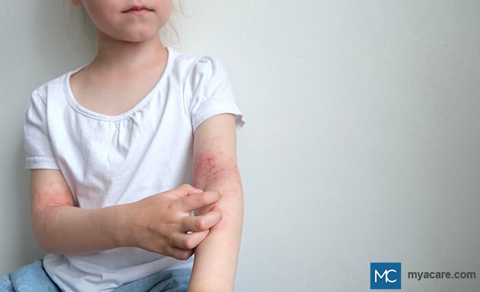
| ATOPIC DERMATITIS
A chronic skin disorder that presents as itchy, red dots, and rashes, Atopic dermatitis (AD) may recur from time to time. Having a personal or family history of asthma, allergic rhinitis, and atopic dermatitis are common factors involved in this skin condition. In infants, AD usually affects the face or cheeks, as well as the extensor surfaces of the arms and legs. In older children and in adults, lesions are most commonly seen in the flexural areas. |
 |
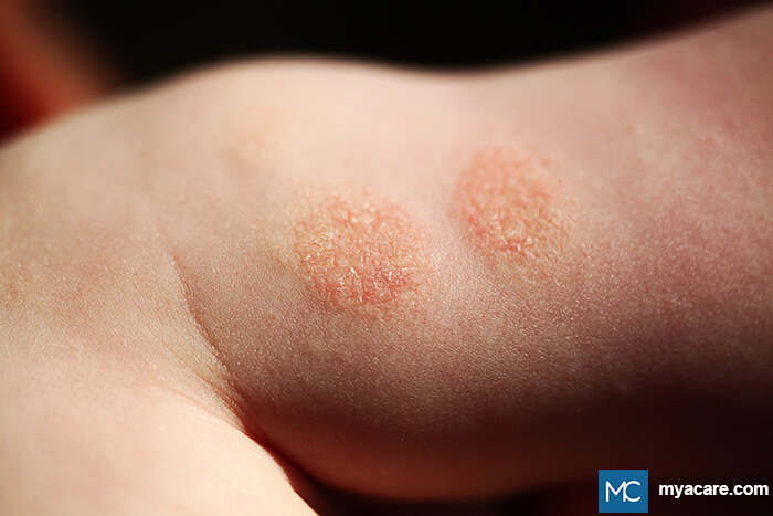
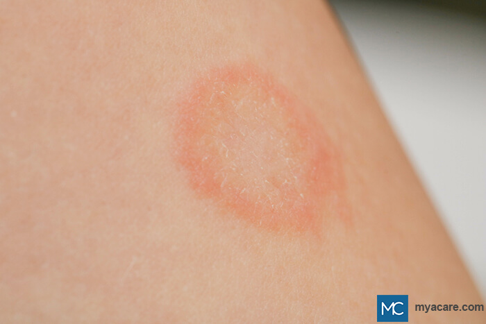
| NUMMULAR ECZEMA
Also called discoid eczema, nummular eczema presents as coin-shaped, itchy, red lesions on the skin that may be oozing or crusted. This usually affects the extremities and may be triggered by a multitude of factors, such as changes in weather and other irritants and allergens. To learn more about Nummular Eczema, please read our blog. |
 |
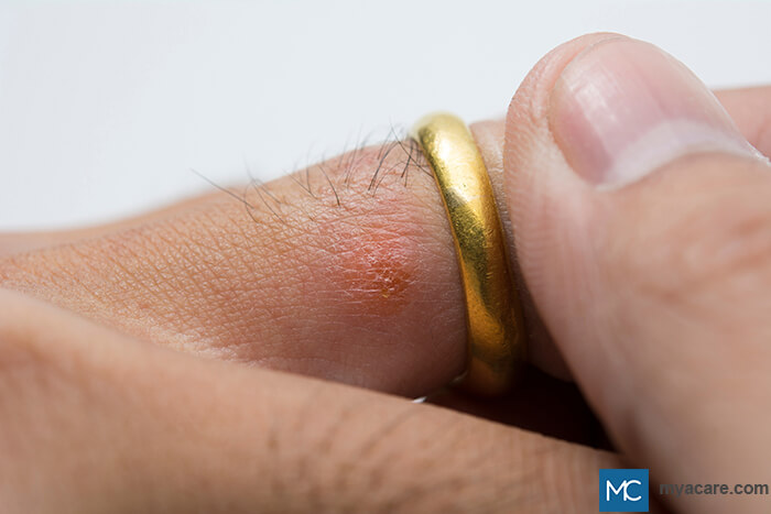
| LICHEN SIMPLEX CHRONICUS AND PRURIGO NODULARIS
Lichen simplex chronicus (LSC) and prurigo nodularis (PN) are skin conditions that are usually caused by persistent and recurring itching, scratching, or rubbing of the skin. They can be due to a systemic disease or by other possible causes, such as having a history of atopic dermatitis. Lesions of LSC and PN are known to have severe itching, triggering the patient to scratch, which causes the thickening of the lesions. LSC lesions are lichenified, scaly, and dry and may be erythematous or hyperpigmented in color. PN, on the other hand, appears as firm nodules. |
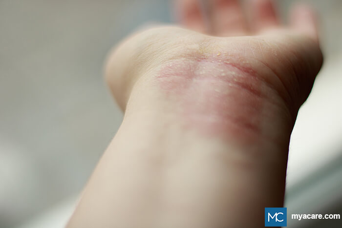 |
| ALLERGIC AND IRRITANT CONTACT DERMATITIS
Allergic contact dermatitis (ACD) and Irritant contact dermatitis (ICD) are skin diseases caused by a reaction to certain allergens that come in contact with the skin. Lesions may appear as itchy red dots or rashes, which can be accompanied by swelling, weeping lesions, or pain, depending on the severity of the disease. |
 |
| SEBORRHEIC DERMATITIS
Seborrheic dermatitis, more commonly known as dandruff, usually appears as red scaly patches on the scalp, face (eyebrows, ears, sides of nose), trunk, and back, especially on the oil-rich areas of the body. Some factors that may cause seborrheic dermatitis include:
To learn more about Seborrheic Dermatitis, please read our blog.
|
 |
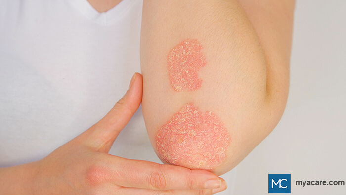
| PSORIASIS
Psoriasis is a chronic skin disorder that is caused by a combination of genetic and environmental factors. Lesions often present as red dots that evolve into thick plaques that have white, scaly surfaces. Lesions usually appear on the scalp and on the extensor surfaces of the skin, such as the elbows and knees, but they can also appear anywhere in the body. To learn more about Psoriasis, please read our blog. |
 |
| PITYRIASIS RUBRA PILARIS
A chronic skin condition, Pityriasis rubra pilaris appears as orange-red scaly plaques that can affect any part of the body but are usually found on the palms and soles. The presence of areas of unaffected skin called “nappes claires” is a diagnostic hallmark. There can also be some nail changes present. To learn more about Pityriasis Rubra Pilaris, please read our blog. |
 |
| PITYRIASIS ROSEA
Pityriasis rosea usually begins with the appearance of a solitary, red, round to oval-shaped, itchy “mother patch” on the trunk. After around two weeks, the mother patch is followed by the appearance of multiple lesions with the same morphology but in smaller sizes. Lesions also have some scaling on the periphery and can form a “Christmas tree” pattern on the upper chest and back. Aside from the trunk, the extremities can also be affected. The etiology is unknown but it is speculated to be viral in nature. Lesions usually disappear in 5-8 weeks. To learn more about Pityriasis Rosea, please read our blog. |
 |
| LICHEN PLANUS
Lesions of lichen planus appear as red-violet, flat-topped, polygonal raised lesions commonly seen on the flexural arms, wrists, and legs. It is associated with fine, white scales called Wickham striae. The cause is unknown, but is postulated to be affected by infectious, immunologic, genetic, and metabolic causes. To learn more about Lichen Planus, please read our blog.
|
 |
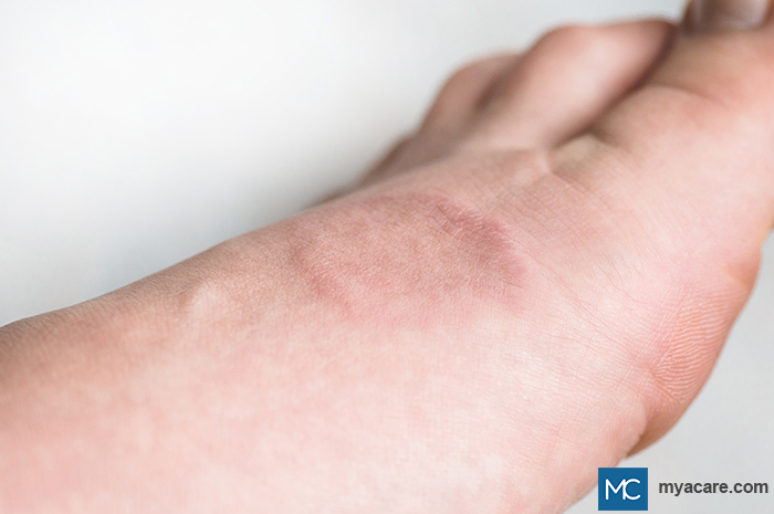
| GRANULOMA ANNULARE
Granuloma annulare is a benign skin disease that presents as annular or ring-like lesions that may be reddish, violaceous, or skin-colored. The most common sites affected include the hands, feet, wrists, ankles, and lower limbs. To learn more about Granuloma Annulare, please read our blog. |
 |
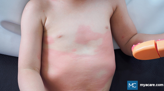
| URTICARIA
Urticaria is a common skin condition that presents as red, itchy bumps or wheals (hives) on the skin. Most cases of urticaria are idiopathic, but some may be caused by infections, chronic autoimmune diseases, and other possible triggers. Lesions are not contagious and usually spontaneously disappear within 24 hours. It affects children, adolescents, and young adults but can still affect older age groups as well. To learn more about Urticaria, please read our blog. |
 |
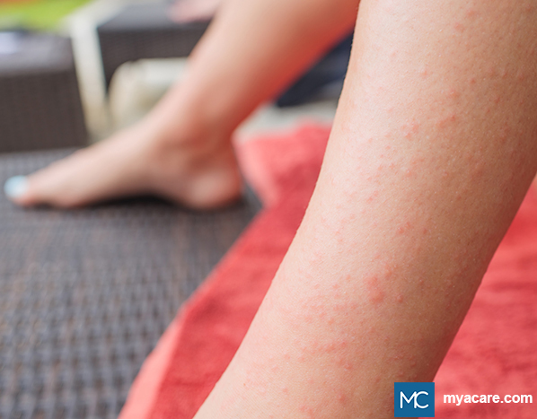
| DRUG HYPERSENSITIVITY REACTIONS
Reactions to drug therapy commonly have skin manifestations and usually present as maculopapular, red, itchy rashes that may be accompanied by other systemic symptoms such as difficulty breathing and swelling of the eyes or lips. Those that present as cutaneous hypersensitivity reactions can also appear with blisters, pustules, or wheals. When hypersensitivity reactions are suspected, it is best to withdraw the offending agent and consult with your doctor as soon as possible. |
 |
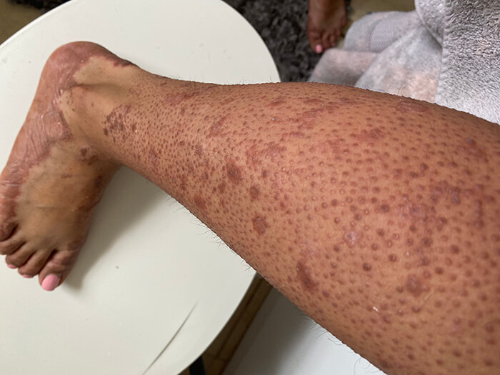
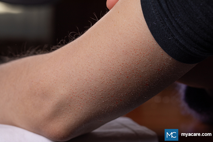
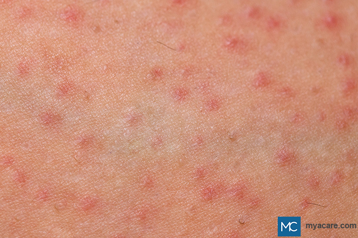
| KERATOSIS PILARIS
Keratosis Pilaris (KP) presents as multiple skin-colored or reddish or light brown bumps most commonly seen on the lateral arms, thighs, or buttocks. Lesions of KP can be itchy or asymptomatic. This is a benign but chronic skin disorder and usually requires no treatment. To learn more about Keratosis Pilaris, please read our blog. |
 |
| PEMPHIGUS VULGARIS
Pemphigus vulgaris is an autoimmune blistering disease that presents as itchy or painful flaccid blisters that can evolve into scaly, crusted erosions on an erythematous base. These can appear anywhere in the body, as well as in mucous membranes. To learn more about Pemphigus Vulgaris, please read our blog. |
 |
| BULLOUS PEMPHIGOID
Bullous Pemphigoid (BP) is another autoimmune blistering disorder that presents with itchy tense blisters on an erythematous base. BP is usually seen in the elderly and is commonly found on the flexural areas, lower abdomen, and thighs, but it can still appear anywhere in the body. These blisters or bullae are usually filled with serous fluid. To learn more about Bullous Pemphigoid, please read our blog.
|
 |
| DERMATITIS HERPETIFORMIS
Dermatitis Herpetiformis appears as vesicles, bumps, or plaques that follow a herpetiform (herpes-like) pattern. DH lesions may be accompanied by itching, burning, or stinging sensation most commonly seen on the elbows, knees, buttocks, shoulders, and sacral areas. The scalp can also be affected. Lesions of DH tend to wax and wane. There is also associated gluten-sensitive enteropathy. To learn more about Dermatitis Herpetiformis, please read our blog. |
 |
| LUPUS ERYTHEMATOSUS
Cutaneous Lupus Erythematosus presents as the classic butterfly rash or malar rash, wherein the lesions appear on the malar eminences and over the bridge of the nose, with sparing of the nasolabial folds. More generalized forms appear as widespread maculopapular rashes on the extensor surfaces of the arms and hands, with sparing of the knuckles. Ultraviolet (UV) radiation is a usual trigger. To learn more about Lupus Erythematosus, please read our blog. |
 |
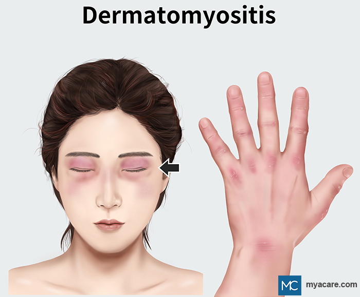
| DERMATOMYOSITIS
Dermatomyositis is an autoimmune disease that presents as reddish to violaceous patches and plaques, which can be found on the scalp, trunk, posterior neck, upper back, and shoulders. These lesions are usually associated with muscle weakness. |
 |
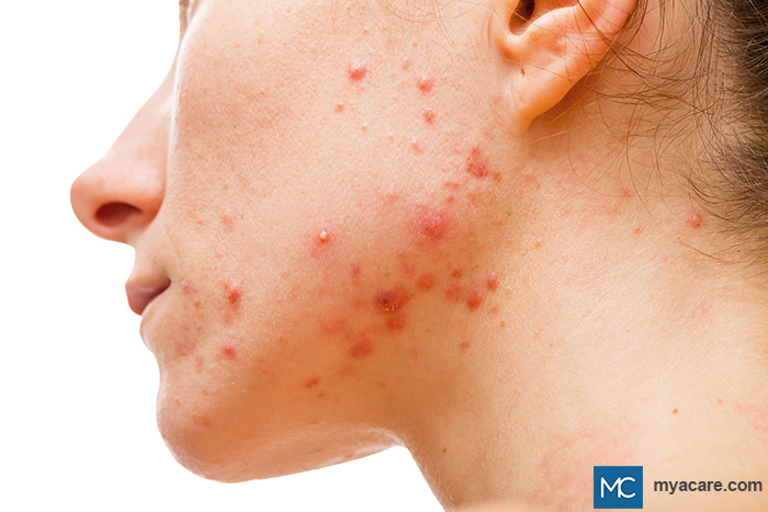
| ACNE VULGARIS
Acne vulgaris is a common chronic inflammatory skin condition that affects the pilosebaceous unit. It is commonly seen in adolescents and presents with various skin lesions such as comedones (whiteheads and blackheads), papules, pustules, and even nodules and cysts in more severe cases. It can affect the face, back, chest, and shoulders. To learn more about Acne vulgaris, please read our blog.
|
 |
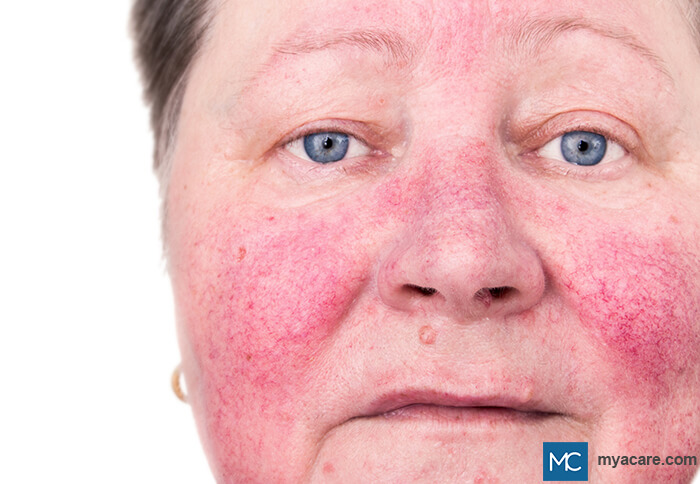
| ROSACEA
Rosacea is another chronic inflammatory skin condition that affects the central facial skin. It is characterized by facial flushing and can present with pimple-like lesions, spider veins or telangiectasias, and phymatous changes in the skin. To learn more about Rosacea, please read our blog. |
 |
| HIDRADENITIS SUPPURATIVA
Hidradenitis suppurative, also known as acne inversa, is a skin disorder that affects the hair follicles and is usually seen in the intertriginous and anogenital areas of the body such as the armpits, breasts, inner thighs, and groin area. Lesions present as tender and painful nodules or lumps that can progress to abscess-like lesions that drain purulent material. To learn more about Hidradenitis Suppurativa, please read our blog. |
 |
| ERYTHEMA TOXICUM NEONATORUM
A skin condition seen in newborns, Erythema toxicum neonatorum presents as lesions that usually appear as blotchy, red patches with central vesicles or pustules. They can affect any part of the body, sparing the palms and soles. It is a benign condition and usually resolves spontaneously in 2-3 weeks. To learn more about Erythema Toxicum Neonatorum, please read our blog.
|
 |
| MILIARIA RUBRA
Miliaria rubra, or “heat rash”, is a skin disorder affecting the eccrine (sweat) glands. Lesions appear as red papules and are usually caused by overheating. |
 |
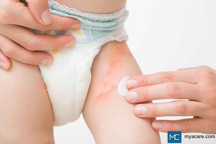
| DIAPER DERMATITIS
Diaper dermatitis is a common skin disorder in infants. Most cases are due to underlying irritant contact dermatitis. Lesions present as red, moist, and sometimes scaly patches on the genital area and buttocks. |
 |
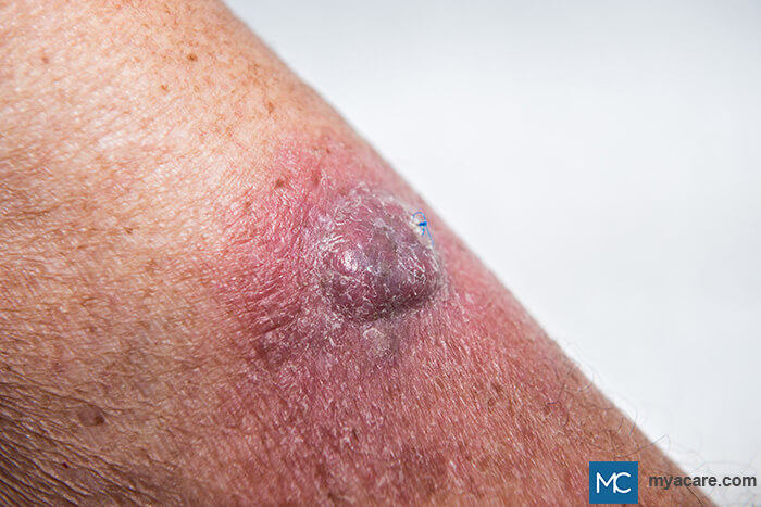
| BASAL CELL CARCINOMA
Basal cell carcinoma (BCC) is the most common skin cancer in humans. There are many types of BCC, but lesions usually appear as papules of nodules that can have ulcerations at the center, as well as telangiectasias and rolled borders. The head and neck are the most common areas affected. To learn more about Basal Cell Carcinoma, please read our blog. |
 |
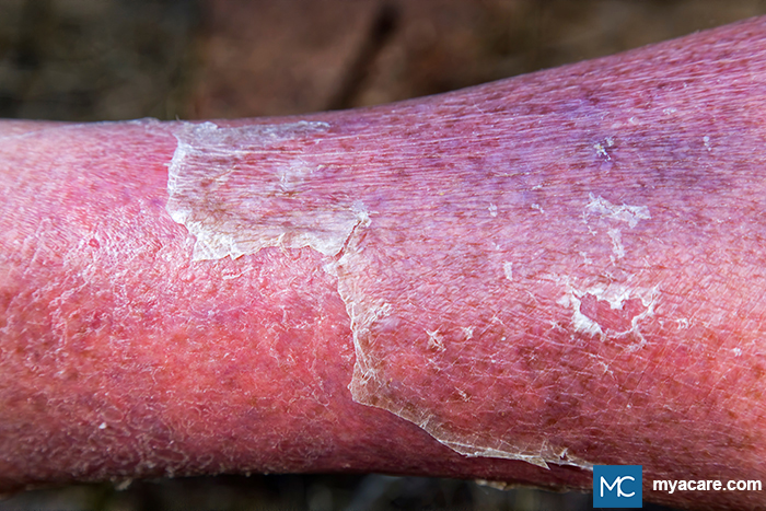
| SQUAMOUS CELL CARCINOMA
Squamous cell carcinoma (SCC) is the second most common skin cancer in humans. There are many risk factors for SCC, but the most important one is high cumulative exposure to UV radiation. Lesions appear as red plaques or nodules that are slowly enlarging. Ulceration is also present. Lesions typically affect sun-exposed areas of the skin, especially the head, face, and neck region. To learn more about Squamous Cell Carcinoma, please read our blog.
|
 |
| INFANTILE HEMANGIOMA
Infantile hemangiomas appear as red birthmarks in infant children. These lesions are benign vascular growths that usually disappear on their own. |
 |
| CUTANEOUS VASCULITIS
Cutaneous vasculitis refers to the skin manifestations of inflamed blood vessels. Lesions usually appear as red spots, which are non-blanching and commonly affect the lower legs. |
 |
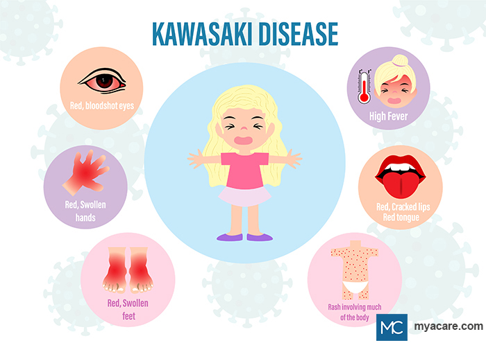
| KAWASAKI DISEASE
Kawasaki disease (KD) is a multisystem disorder that usually affects young children. This disease causes inflammation of the blood vessels. Skin lesions appear as red, itchy morbilliform rashes on the trunk and extremities, usually can be accompanied by fever, mucosal changes such as red lips or strawberry tongue, cervical lymphadenopathy, conjunctival injections, and swelling of the hands and feet. To learn more about Kawasaki disease (KD), please read our blog. |
 |
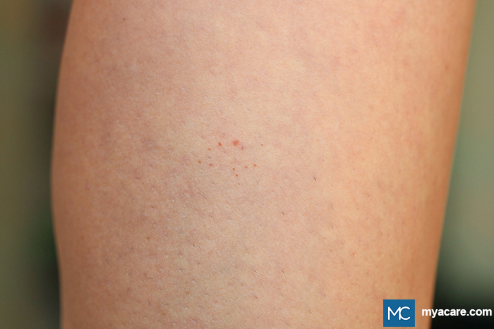
| PIGMENTED PURPURIC DERMATOSES
Pigmented Purpuric Dermatoses (PPD) are lesions that appear as reddish-brown patches or dots, associated with pigmentation and, sometimes, telangiectasias. They are often caused by leaky capillaries. The most affected areas are the lower legs. |
 |
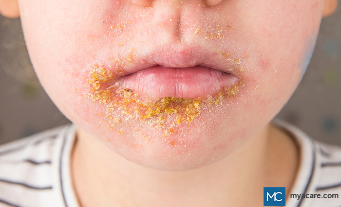
| IMPETIGO
Impetigo is a superficial bacterial skin infection presenting as red papules or fluid-filled vesicles that can rupture and form plaques with honey-colored crusts commonly found on the face or extremities, especially on the area of the nose or lips. Impetigo frequently affects young children. To learn more about Impetigo, please read our blog. |
 |
| FOLLICULITIS
Folliculitis is another bacterial skin infection that specifically affects the hair follicles. Lesions appear as small, dome-shaped pustules with redness and some swelling around the hair follicle openings. Lesions can be itchy and are usually seen on hair-bearing areas of the body, such as the armpits, extremities, and buttocks. |
 |
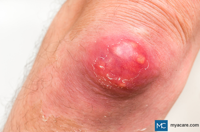
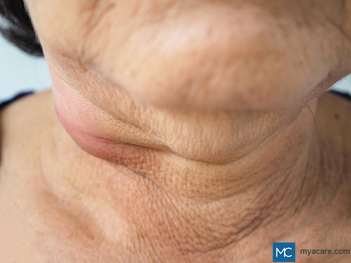
| FURUNCLES AND CARBUNCLES
Furuncles and carbuncles are deeper infections of the hair follicles. Furuncles, or boils, commonly appear as red, tender nodules which can affect the neck, face, armpits, buttocks, and any other hair-bearing areas of the body. When furuncles cluster together, they form a carbuncle. To learn more about Furuncles and Carbuncles, please read our blog. |
 |
| ERYTHRASMA
Erythrasma is a superficial bacterial infection characterized by reddish-brown patches that are usually found on the intertriginous areas of the body. Diagnosis is usually aided by the presence of coral red fluorescence upon examination with a Wood’s lamp. |
 |
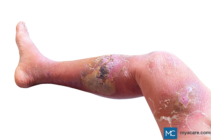
| CELLULITIS
Cellulitis is a common bacterial infection affecting the deep dermis and subcutaneous tissue. Lesions are red and painful, can have accompanying edema or swelling, and are warm-to-touch. The lower extremities are most commonly affected. To learn more about Cellulitis, please read our blog. |
 |
| ERYSIPELAS
Erysipelas is commonly mistaken for cellulitis, as it appears as red plaques and can have accompanying edema. The differentiating factor is that lesions of erysipelas have sharply marginated and well-defined borders compared to the ill-defined borders of cellulitis. Most lesions affect the legs but can also appear on the face and upper extremities. |
 |
| CUTANEOUS TUBERCULOSIS
Tuberculosis (TB) does not only affect the lungs but can also affect the skin. Scrofuloderma and Lupus Vulgaris (LV) are some of the few kinds of cutaneous TB. Scrofuloderma lesions are commonly found in the submandibular, parotid, or supraclavicular area. LV lesions are usually seen on the head and neck. To learn more about Cutaneous Tuberculosis (CTB), please read our blog. |
 |
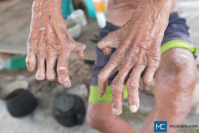
| LEPROSY
Leprosy, or Hansen’s Disease, is a mycobacterial infection that affects the skin, peripheral nerves, nasal mucosa, and eyes. Skin lesions of leprosy appear as red, hypopigmented, or hyperpigmented patches or plaques that have impaired or no sensation. There may also be nodules or lumps on the face, earlobes, or any part of the skin, as well as accompanying muscle weakness, nerve enlargement, or eye problems. Disfigurement of hands and feet is also possible. To learn more about Leprosy, please read our blog. |
 |
| SUPERFICIAL FUNGAL INFECTIONS
Superficial fungal infections can affect the hair, skin, and nails. Lesions usually appear as red itchy patches and plaques that tend to have advancing borders and scales. To learn more about Superficial Fungal Infections, please read our blog.
|
 |
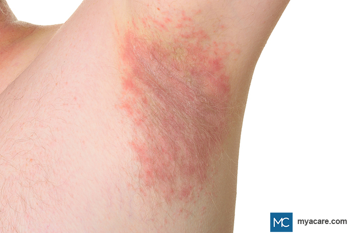
| CANDIDIASIS
Candidiasis is also a fungal infection that presents as beefy-red patches and plaques with satellite papules, usually affecting the intertriginous areas such as the armpits, groin folds, and inframammary area. To learn more about Candidiasis, please read our blog.
|
 |
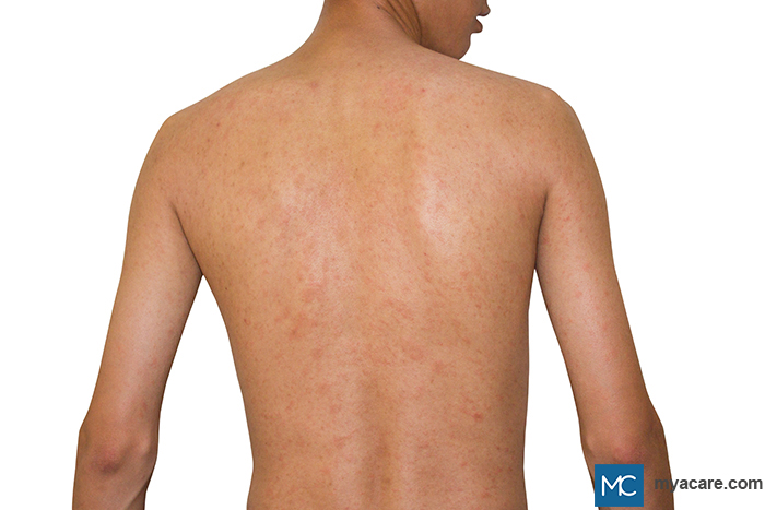
| MEASLES
Measles, otherwise known as rubeola, is a viral infection that presents as small, nonpruritic, red dots or macules that start from the head or face and then spread downwards to the trunk and extremities. Lesions are usually preceded by fever, body weakness, colds, and cough. “Koplik’s spots” are also present on the oral mucosa. These are small, red macules that have a white speck on them. To learn more about Measles, please read our blog. |
 |
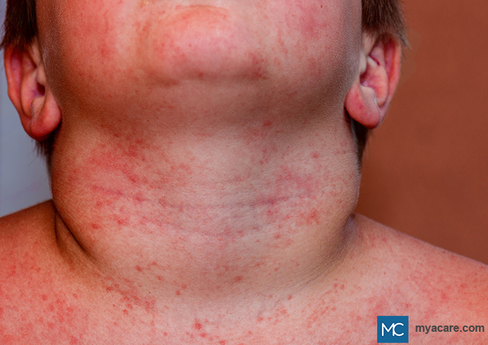
| RUBELLA
Rubella, also called German measles or 3-day measles, is a viral condition characterized by the presence of itchy pink to red dots that begin on the face and then increase to involve the neck, trunk, and extremities. Lesions can be associated with fever, muscle pains, sore throat, cough, and enlarged lymph nodes. |
 |
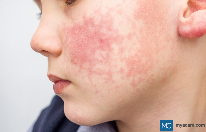
| ERYTHEMA INFECTIOSUM
Erythema infectiosum, or Fifth disease, is a viral skin disease that commonly occurs in children. It presents with bright-red lesions on the malar surfaces, giving a “slapped-cheek” appearance on the face, followed by a lace-like eruption of lesions on the trunk and extremities. There may be accompanying symptoms such as headache, low-grade fever, body weakness, and joint pains. |
 |
| HAND-FOOT-MOUTH DISEASE
Hand-Foot-Mouth disease is a viral skin condition that initially presents with low-grade fever and body weakness. The lesions can appear as bright pink dots and raised lesions on the palms and soles accompanied by oral lesions as well. To learn more about Hand-Foot-Mouth Disease, please read our blog.
|
 |
| VARICELLA
Varicella, or chickenpox, is a highly contagious viral condition caused by the varicella zoster virus. Varicella lesions appear as vesicles or fluid-filled small blisters on a red base, usually starting on the face and scalp and spreading to the trunk. Mild fever, body weakness, and headache are some of the accompanying symptoms. |
 |
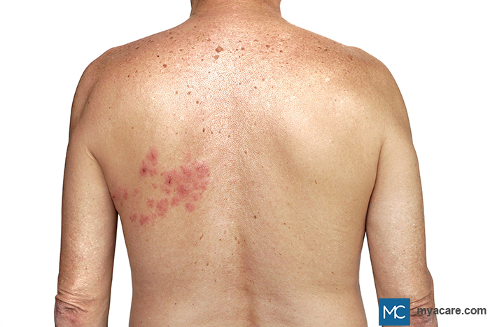
| HERPES ZOSTER
Herpes zoster or shingles is also caused by the varicella virus. Lesions also appear as fluid-filled small blisters that are clustered together, usually on one side of the body. Lesions are associated with pain. To learn more about Herpes zoster, please read our blog. |
 |
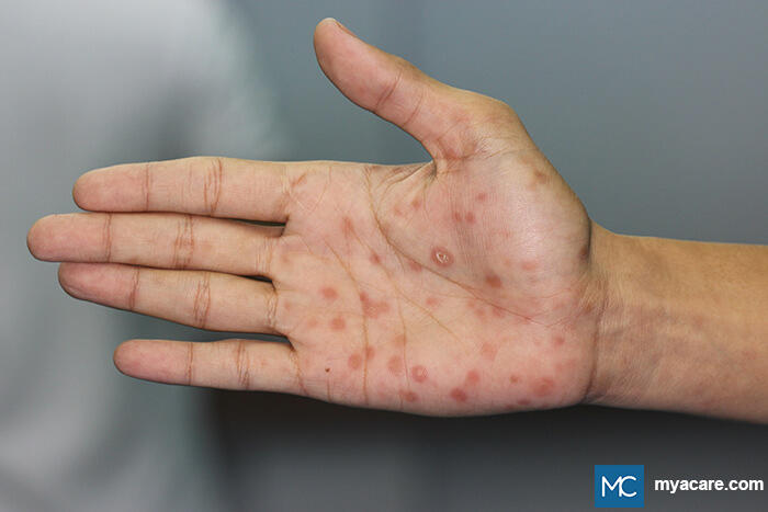
| SECONDARY SYPHILIS
Secondary Syphilis is an infectious disease and is mainly sexually transmitted. There are 4 phases of the disease, and cutaneous manifestations are typically seen in the secondary stage. Lesions appear as red to copper-colored macules or papules that are sometimes scaly and usually not itchy. Lesions appear on the palms and soles but can usually affect the other parts of the body as well. To learn more about Secondary Syphilis, please read our blog. |
 |
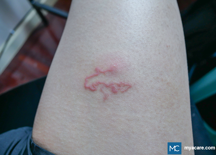
| CUTANEOUS LARVA MIGRANS
Lesions of cutaneous larva migrans appear as very itchy, serpiginous, and linear lesions usually caused by larval migration under the skin. To learn more about Cutaneous Larva Migrans, please read our blog.
|
 |
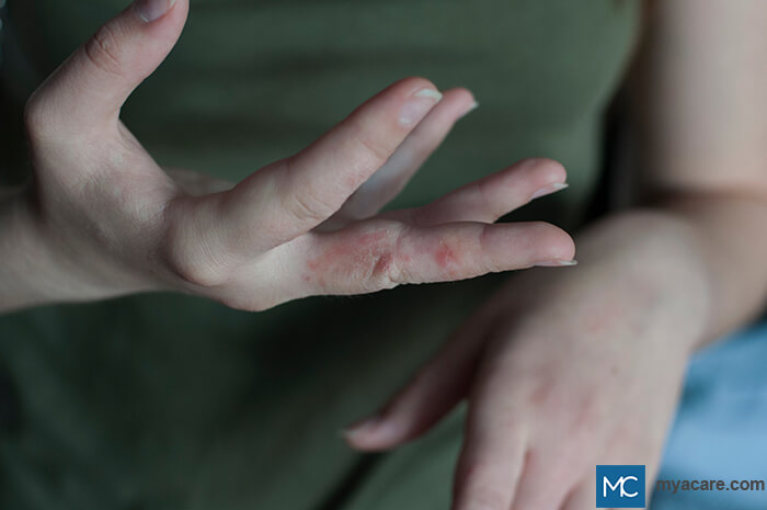
| SCABIES
Scabies is a very common infestation seen in many different parts of the world. Transmission is due to the scabies mite being transferred either by direct contact or fomite transmission. Lesions usually appear as red dots and papules that are usually very itchy at night. It affects the armpits, wrists, abdomen, inguinal area, and other parts of the body. It is common for many members of the same household to exhibit the same lesions. To learn more about Scabies, please read our blog. |
 |
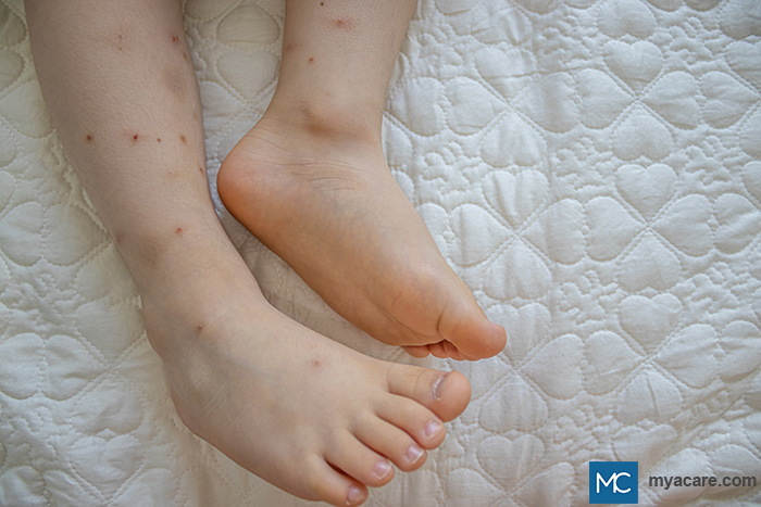
| ARTHROPOD BITE REACTION
Many arthropods or insects can bite or sting humans and cause skin reactions. Bedbug bites are one of the most common skin conditions due to arthropods. Lesions usually appear as a row of 3 or more red, itchy bites, called the “breakfast, lunch, and dinner” sign. |
 |
There are many skin disorders that can appear as red rashes or red dots. It is best to consult a dermatologist so that the correct diagnosis is achieved and the proper treatment is given.
To search for the best health providers for dermatology in Croatia, Germany, Greece, Italy, Malaysia, Singapore, Slovakia Spain, Thailand, The UAE, the UK, and the US, please use our free search engine.
To search for the best healthcare providers worldwide, please use the Mya Care search engine.
Dr. Lauren Livelo is a board-certified dermatologist from the Philippines. She has a degree in Medicine from the University of the East Ramon Magsaysay Memorial Medical Center, and has completed her dermatology residency training in the Research Institute for Tropical Medicine. Aside from her private practice, she enjoys writing about skin care and diseases of the skin.
References:
Featured Blogs

Medically Induced Coma: What It Is, How It Works, Who Benefits, and Recovery Outcomes

